
Through the Smoke and Mirrors: Thoracolumbar Hemilaminectomy (T13-L1) and Complex Spinal Deformity in a Paraparetic French Bulldog
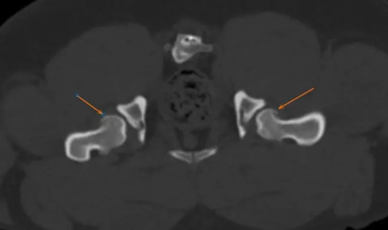
When an owner rushes a French Bulldog into my consultation room complaining of "sudden hindlimb weakness," the easiest mistake in veterinary medicine is to blame the hips. This Frenchie had already been diagnosed with hip disease at an outside clinic, and his radiographs did indeed show hip dysplasia. But as I remind colleagues repeatedly: hip joints do not cause acute neurological paralysis! Neurological examination at Guangzhou Boss Animal Hospital confirmed loss of conscious proprioception, reflex abnormalities, and severe paraparesis. High-resolution CT cut through the anatomical confusion, identifying a classic diagnostic trap: while the dog had congenital hemivertebrae at T4 and T8 alongside shallow hips, the real emergency was an acute T13-L1 intervertebral disc herniation compressing the spinal cord by 30–40%. I performed an emergency T13-L1 hemilaminectomy to decompress the cord. True to the resilient nature of the breed, he woke up from spinal surgery and immediately demanded his food bowl. Within weeks, he was back on his feet, walking soundly.
Total Knee Arthroplasty (TKA) in a 41 kg Doberman Pinscher: Overcoming End-Stage Osteoarthritis with a Biological Dual-Mobility System
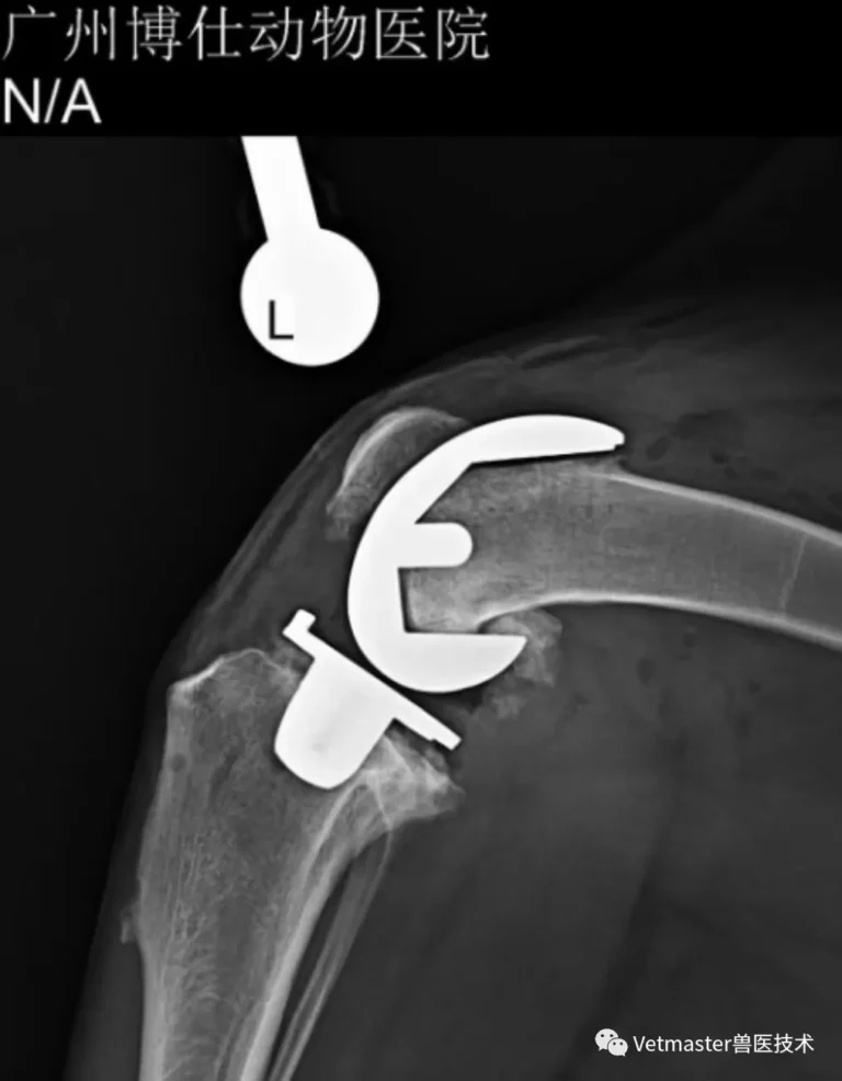
When an owner drives hundreds of miles from outside the province to bring a 41 kg adult male Doberman Pinscher to Guangzhou Boss Animal Hospital, you know you are dealing with a severe, chronic crisis. The dog had suffered from recurrent bouts of severe knee swelling and lameness that temporarily waxed and waned, masking an insidious, chronic cranial cruciate ligament rupture. By the time he reached my operating table, the left stifle was in end-stage collapse—encased in massive osteophytes with complete destruction of both the femorotibial and patellofemoral articular cartilage. In a destroyed joint like this, standard TPLO is useless because it cannot heal eburnated, grinding bone. Using our VVV digital surgical planning software, I mapped out the reconstruction and implanted a Size 40 Vetmaster biological dual-mobility total knee replacement. Despite dense fibrous joint contractures that made joint space distraction an intense physical and technical battle, we achieved textbook alignment with an exact 12° posterior tibial slope. Today, this massive working dog has regained painless, powerful athletic function.
Swiss KYON Patellar Groove Replacement (PGR) in a Pomeranian: When Conservative Delay Destroys the Trochlea
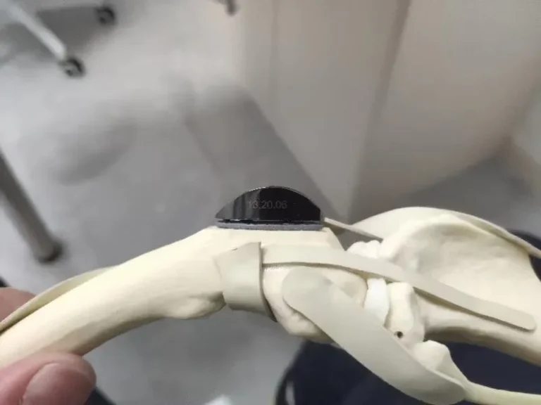
Many pet owners believe patellar luxation can be managed indefinitely with supplements and rest. This case of a 4-year-old, 5.5 kg male Pomeranian proves the harsh reality of that misconception. Having deferred surgery for over a year at an outside clinic, the dog was brought to Guangzhou Boss Animal Hospital with marked left hindlimb lameness. Physical examination revealed Grade 3 medial patellar luxation accompanied by harsh, bone-on-bone joint crepitus. Radiographs confirmed what my fingers felt: the medial trochlear ridge was flattened, and the articular cartilage was worn down to eburnated subchondral bone. In this end-stage scenario, traditional trochlear deepening is useless—carving into a joint that has no cartilage left only forces the patella to grind on raw bone. To restore smooth mechanics, I performed a Patellar Groove Replacement (PGR) using a Swiss KYON Size 2 prosthesis. Today, the patella tracks flawlessly over the polished artificial sulcus without pain, underscoring my golden rule: native living cartilage is always better than metal, but an engineered prosthesis is infinitely superior to chronic bone-on-bone destruction.
Preserving the Native Joint: Double Pelvic Osteotomy (DPO) in a 6-Month-Old Golden Retriever with Hip Dysplasia
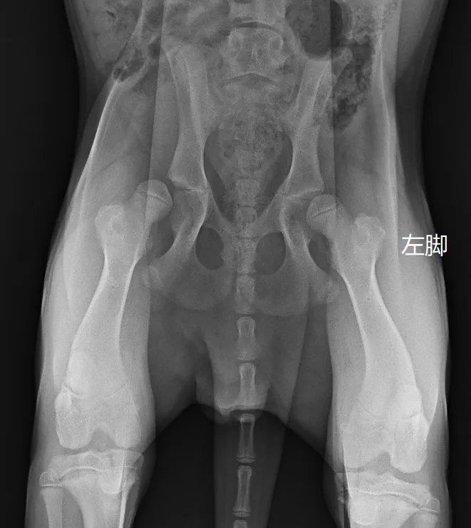
Many in the veterinary community know me as someone who performs a high volume of total hip replacements. But total hip replacement is ultimately a salvage procedure—we replace a joint only when the native cartilage is irrevocably destroyed. My true clinical priority is always joint preservation: saving the animal's own, natural hip whenever possible. In this case, a 6-month-and-24-day-old Golden Retriever (23 kg) presented with abnormal hindlimb ambulation. Comprehensive screening under sedation revealed significant bilateral laxity. The right hip had extreme laxity (DI 1.03), ruling out osteotomy, but the left hip (DI 0.71, subluxation angle 18°, reduction angle 37°) sat squarely within the golden window for Double Pelvic Osteotomy (DPO). By performing a DPO on the left pelvis, I rotated the acetabulum to dramatically increase dorsal coverage over the growing femoral head. At the two-month follow-up, the dog's left thigh muscle diameter was 20% larger than the non-operated right side, demonstrating restored weight-bearing and proving the immense power of early proactive joint preservation.