
Osteotomy & Joint Surgery
DPO/JPS early intervention, patellar luxation, PGR, and cruciate ligament osteotomies.
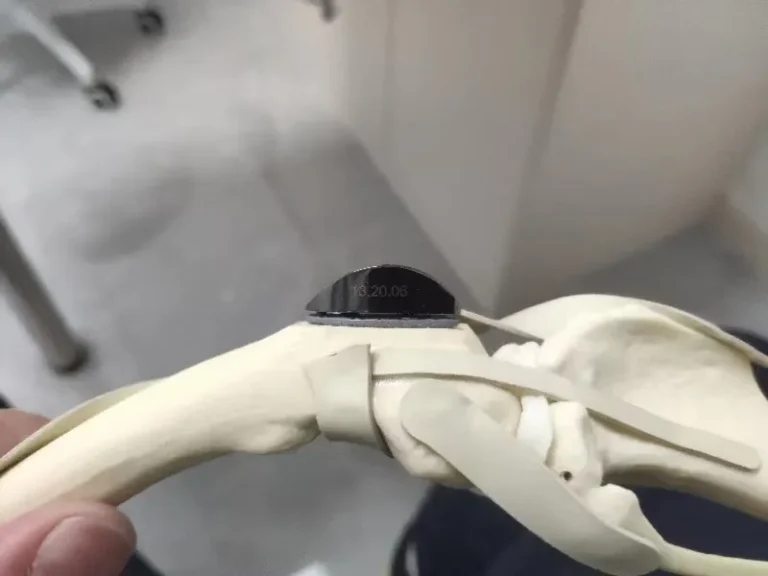
Swiss KYON Patellar Groove Replacement (PGR) in a Pomeranian: When Conservative Delay Destroys the Trochlea
Many pet owners believe patellar luxation can be managed indefinitely with supplements and rest. This case of a 4-year-old, 5.5 kg male Pomeranian proves the harsh reality of that misconception. Having deferred surgery for over a year at an outside clinic, the dog was brought to Guangzhou Boss Animal Hospital with marked left hindlimb lameness. Physical examination revealed Grade 3 medial patellar luxation accompanied by harsh, bone-on-bone joint crepitus. Radiographs confirmed what my fingers felt: the medial trochlear ridge was flattened, and the articular cartilage was worn down to eburnated subchondral bone. In this end-stage scenario, traditional trochlear deepening is useless—carving into a joint that has no cartilage left only forces the patella to grind on raw bone. To restore smooth mechanics, I performed a Patellar Groove Replacement (PGR) using a Swiss KYON Size 2 prosthesis. Today, the patella tracks flawlessly over the polished artificial sulcus without pain, underscoring my golden rule: native living cartilage is always better than metal, but an engineered prosthesis is infinitely superior to chronic bone-on-bone destruction.
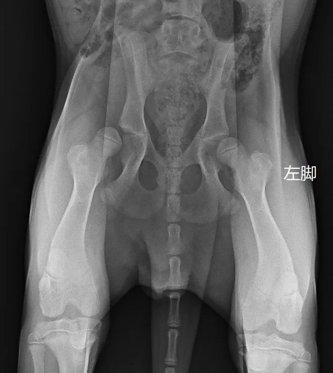
Preserving the Native Joint: Double Pelvic Osteotomy (DPO) in a 6-Month-Old Golden Retriever with Hip Dysplasia
Many in the veterinary community know me as someone who performs a high volume of total hip replacements. But total hip replacement is ultimately a salvage procedure—we replace a joint only when the native cartilage is irrevocably destroyed. My true clinical priority is always joint preservation: saving the animal's own, natural hip whenever possible. In this case, a 6-month-and-24-day-old Golden Retriever (23 kg) presented with abnormal hindlimb ambulation. Comprehensive screening under sedation revealed significant bilateral laxity. The right hip had extreme laxity (DI 1.03), ruling out osteotomy, but the left hip (DI 0.71, subluxation angle 18°, reduction angle 37°) sat squarely within the golden window for Double Pelvic Osteotomy (DPO). By performing a DPO on the left pelvis, I rotated the acetabulum to dramatically increase dorsal coverage over the growing femoral head. At the two-month follow-up, the dog's left thigh muscle diameter was 20% larger than the non-operated right side, demonstrating restored weight-bearing and proving the immense power of early proactive joint preservation.