
Salvaging a Disaster: Revision of a Four-Times Luxated Total Hip Replacement in a Border Collie Using a Dual-Mobility System
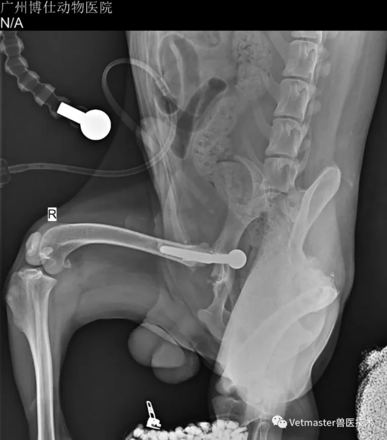
When this desperate owner drove over 1,000 kilometers to bring his 5.5-year-old, 22.5 kg Border Collie to Guangzhou Boss Animal Hospital, the dog was facing his fifth hip surgery in just over two months. Following an initial hybrid replacement at another clinic, the dog had suffered four consecutive dorsal hip luxations, each recurring within two to five days despite repeated attempts to alter cup angles down to an extreme 25°. Analyzing the patient's CT, I identified a striking biomechanical contradiction: an overly closed cup should cause ventral luxation, yet this dog kept popping out dorsally—proving the dorsal muscular and capsular envelope had been completely destroyed. My colleague Dr. Yu Jiezhou and I revised the joint using the Vetmaster cementless dual-mobility system reinforced with synthetic ligament suspension. By replacing mechanical friction with dual-mobility jump distance, we achieved rock-solid stability—demonstrated intraoperatively by gently lifting the dog off the table by the femoral neck without dislocation. Today, the dog walks soundly, free from the revision nightmare.
Limb Salvage with a Custom "Super-Alloy" Bionic Prosthesis in a Dog with Radial Nerve Paralysis: Susie’s Story
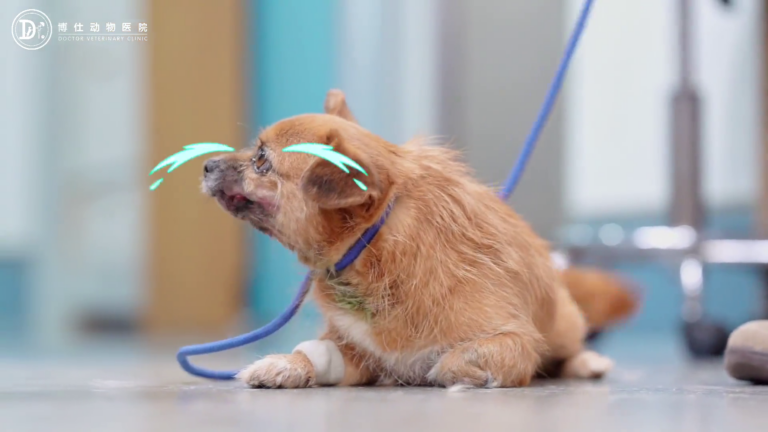
In clinical practice, canine radial nerve paralysis almost always ends with the same grim verdict: high-limb amputation. When the radial nerve fails, the dog loses extensor tone, leaving the paw to drag and knuckle across the ground until chronic friction causes relentless ulceration and deep infection. That was the exact crossroads facing Susie, a little dog brought to me after being scheduled for amputation. Unwilling to accept losing her forelimb if a viable engineering alternative existed, I designed and implanted a custom "super-alloy" bionic endo-exo prosthesis tailored specifically to her skeletal anatomy. Today, Susie is walking comfortably on her new bionic leg, free of pain and infection, proving that customized bionic limb salvage is a realistic, life-changing reality in small animal surgery.
Sequential Bilateral Biological Total Hip Replacement in a British Shorthair Cat: Anatomy, Technique, and Overcoming Contralateral Artifacts
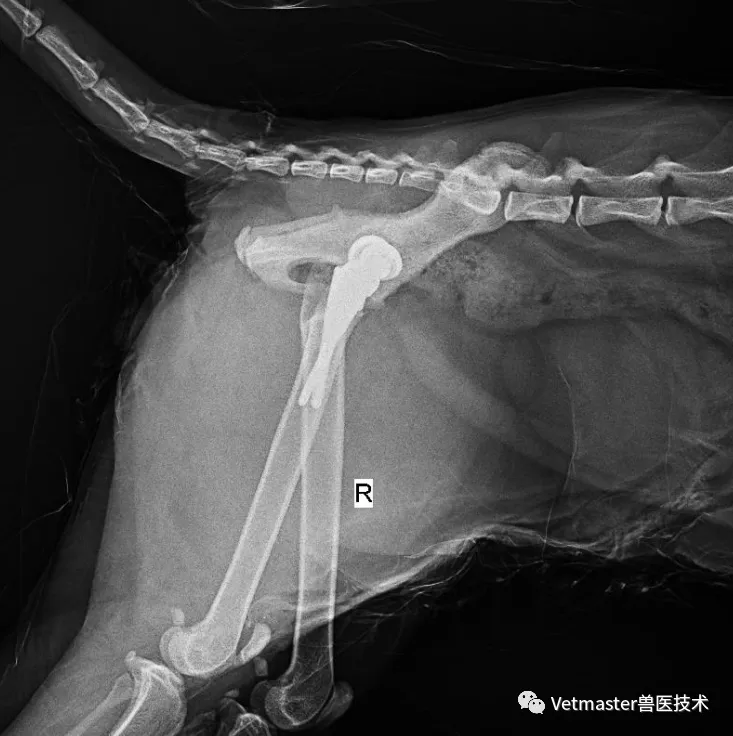
Feline hip dysplasia is vastly underdiagnosed because cats are masters at masking chronic orthopedic pain. In this case, a 2-year-old male British Shorthair weighing 5.8 kg presented with severe bilateral hip laxity, advanced articular wear, and marked lameness. Cats possess unique anatomy: an exceptionally tiny acetabulum paired with a straight, "stovepipe" femoral canal, along with explosive jumping biomechanics requiring extreme range of motion. To restore lifelong, pain-free athletic ability without dislocation, I performed sequential bilateral cementless total hip replacements (THR) using the Vetmaster dual-mobility system (12 mm cup, #3 stem). This report details the technical execution of both stages—including the specific intraoperative challenge during the second stage, where the contralateral metal prosthesis blocked lateral fluoroscopic cup measurement, requiring precise C-arm patient leveling and mechanical alignment guidance.
Custom 3D-Printed Biological Total Knee Arthroplasty (TKA) in a Dog: Clinical Case and 2.5-Month Outcome
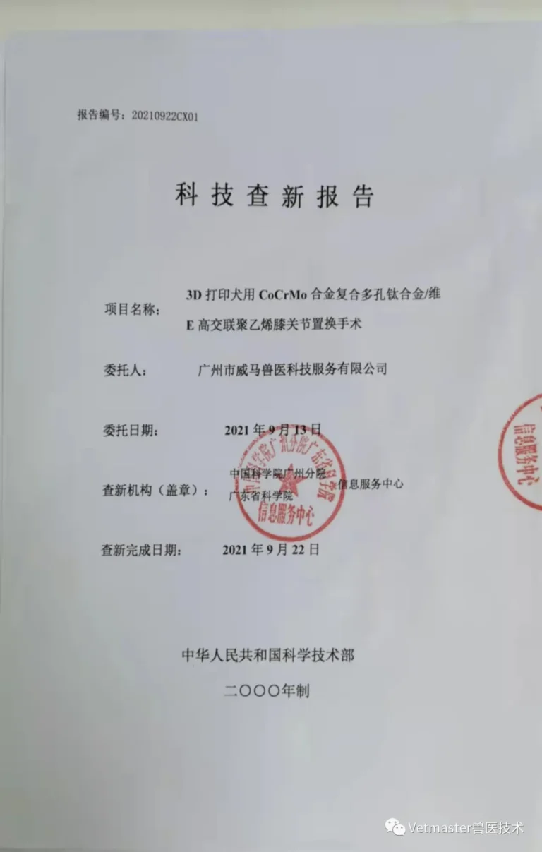
In this case, I document what was the first reported biological canine total knee arthroplasty (TKA) using custom 3D-printed implants and disposable patient-matched instrumentation in China. The patient, a 25 kg Husky, suffered from end-stage osteoarthritis and severe meniscal damage triggered by chronic cranial cruciate ligament rupture. Facing a complete lack of off-the-shelf knee prostheses suited to this dog, my surgical team at Guangzhou Boss Animal Hospital collaborated with Associate Professor Zhang Jun and the materials engineering team at the Guangdong Academy of Sciences. Together, we designed and 3D-printed a cementless CoCrMo/porous titanium/Vitamin E-stabilized polyethylene implant system. Following 1:1 benchtop simulation, we successfully placed the prosthesis. By day one post-op, the dog was weight-bearing without pain; at 2.5 months, he was galloping freely off-leash across the grass.