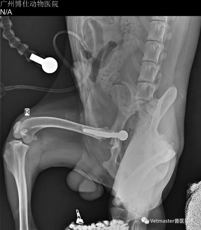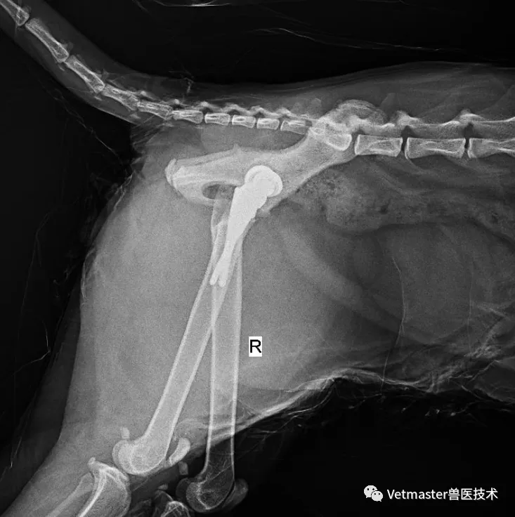
1. The R&D Journey: Bringing Biological Knee Replacement to Chinese Veterinary Surgery
Canine total knee arthroplasty (TKA) is one of the most technically unforgiving summits in small animal orthopedics. For years, domestic veterinary medicine had virtually no answer for a dog whose knee was structurally ruined beyond the reach of osteotomies. The specialized instrumentation and implants were held exclusively by a handful of Western companies, carrying astronomical costs and no commercial availability in China.
Years ago, my team and I decided that Chinese veterinary medicine needed its own independent, world-class answer. Led by my colleague Mr. Ye Cundong (veterinary orthopedic researcher at Guangdong AIB Vocational College and PhD candidate at South China Agricultural University), and developed in close collaboration with Associate Professor Zhang Jun, our materials engineering partners at the Guangdong Academy of Sciences, and under the academic guidance of Professor Li Shoujun's surgical team at SCAU, we engineered the Vetmaster cementless dual-mobility total knee replacement system.
Applying biological dual-mobility principles to canine total knee arthroplasty represents an authentic, pioneering breakthrough. It was designed from scratch to deliver robust multi-planar stability, immediate press-fit osseointegration, and maximum resistance against postoperative subluxation.
Cases like this 41 kg Doberman Pinscher were precisely why we spent years developing it.
2. Case Presentation: A 41 kg Heavyweight in Chronic Collapse
The patient was a 6-year-old intact male Doberman Pinscher weighing a muscular 41 kg. His owners drove all the way from another province to seek a definitive solution at Guangzhou Boss Animal Hospital.
His clinical history was classic for undiagnosed, chronic cranial cruciate ligament (CCL) pathology:
- For months, the dog had suffered recurrent episodes of left stifle swelling and severe lameness.
- Because each episode partially resolved after rest and medication, the owners had assumed it was a series of minor sprains.
- In reality, the cruciate ligament had experienced a progressive, chronic tear. The persistent micro-instability had acted like an abrasive grinding stone, destroying the joint day after day until conservative therapy completely collapsed.
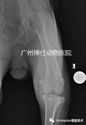
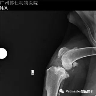
Physical palpation under sedation revealed gross stifle enlargement, dense periarticular fibrous thickening, severe pain on manipulation, and marked loss of flexion. Digital radiographs were alarming: the entire joint was engulfed in massive, florid osteophytosis spanning both femoral condyles, the tibial plateau, and the patellar margins. Orthogonal screening of the hips showed clean, healthy sockets, confirming that his debilitating lameness was 100% localized to this destroyed left stifle.
3. The Clinical Crossroads: Why TPLO Was Off the Table
In everyday clinical practice, many veterinarians instinctively schedule any cruciate-deficient dog for a Tibial Plateau Leveling Osteotomy (TPLO). But as an orthopedic surgeon, you must know where an osteotomy’s therapeutic boundaries end.
- The Fallacy of TPLO in End-Stage DJD: TPLO is a brilliant mechanical procedure designed to neutralize cranial tibial thrust by altering the tibial plateau slope. But TPLO relies on having viable joint cartilage left to glide on. It does nothing to repair or replace denuded, eburnated bone. In a knee where full-thickness cartilage loss has exposed bare subchondral bone across all compartments, performing a TPLO is an exercise in futility. The joint will stop thrusting, but it will continue to grind bone on bone, leaving the dog in chronic, low-grade agony.
- Why TPLO + PGR Falls Short: Combining a TPLO with a Patellar Groove Replacement (PGR) can resurface the trochlea, but it completely ignores the destroyed femorotibial articulation between the condyles and the tibial plateau.
When degenerative joint disease reaches end-stage collapse, there is only one definitive cure that completely eliminates joint pain: Total Knee Arthroplasty (TKA), replacing every worn friction surface in the stifle joint.
4. Digital Planning and the Intraoperative Battlefield
Operating on a 41 kg Doberman with chronic stifle pathology is an immense physical and surgical challenge.
- Digital Preoperative Templating (VVV Software):
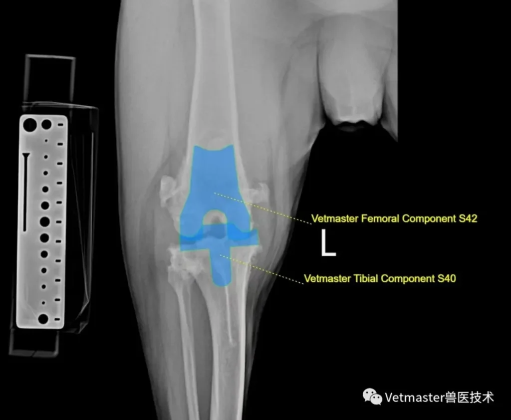
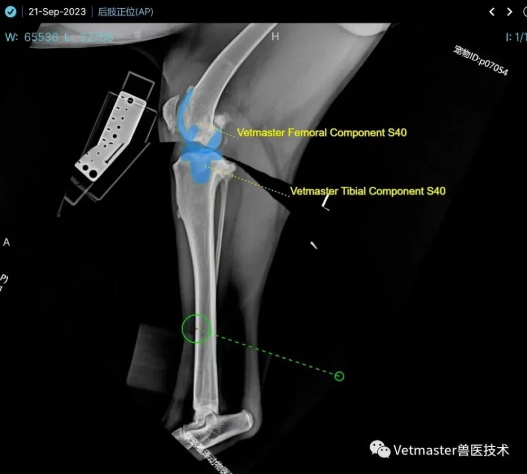
Using our specialized VVV orthopedic planning platform, I calibrated the patient's orthogonal radiographic and CT datasets. Because the osteophytes distorted the natural anatomical landmarks, digital templating was vital to calculate our bone cut depths and re-establish the true joint line. We selected a Size 40 Vetmaster biological knee prosthesis.
- The Soft-Tissue Nightmare: Severe Contracture:
Chronic inflammation in a large, heavily muscled dog creates an intraoperative battlefield. The joint capsule was leathery and millimeters thick; dense fibrosis encased the collateral ligaments. The single hardest part of this surgery was achieving adequate joint space distraction. Without releasing contractures, you cannot insert your cutting blocks or inspect the posterior compartments without tearing the collateral ligaments. - Intraoperative Pathology:
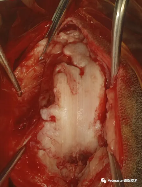
Once the joint was exposed, the native anatomy was almost unrecognizable. The cruciate ligament was completely torn, the menisci were shredded into fibrous remnants, and the femoral trochlea was buried beneath massive osteophytes.
- Precision Bone Resection & Biological Seating:
Working methodically with our specialized Vetmaster single-use cutting guides, I executed the distal and chamfer cuts on the femoral condyles and performed the proximal tibial plateau resection. The biological Size 40 femoral and tibial trays were driven home into vascularized cancellous bone, achieving an immediate, rock-solid press-fit. We inserted the mobile dual-mobility meniscal bearing insert and reduced the joint.
5. Postoperative Validation and Recovery
Postoperative orthogonal radiographs and multi-planar CT reconstructions confirmed exceptional execution:
- The femoral condylar component matched the distal femoral cortices seamlessly.
- The tibial plateau component was seated dead-center, maintaining a pristine 12-degree posterior slope, perfectly balancing flexion-extension stability with quadriceps efficiency.
- CT 3D volumetric rendering verified flush cortical seating with zero overhang or rotational malalignment.
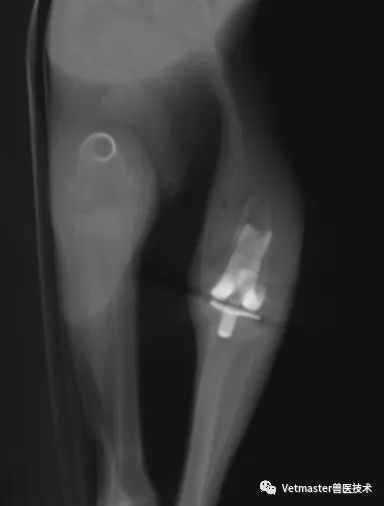
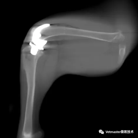
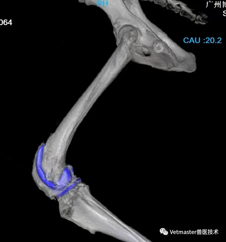
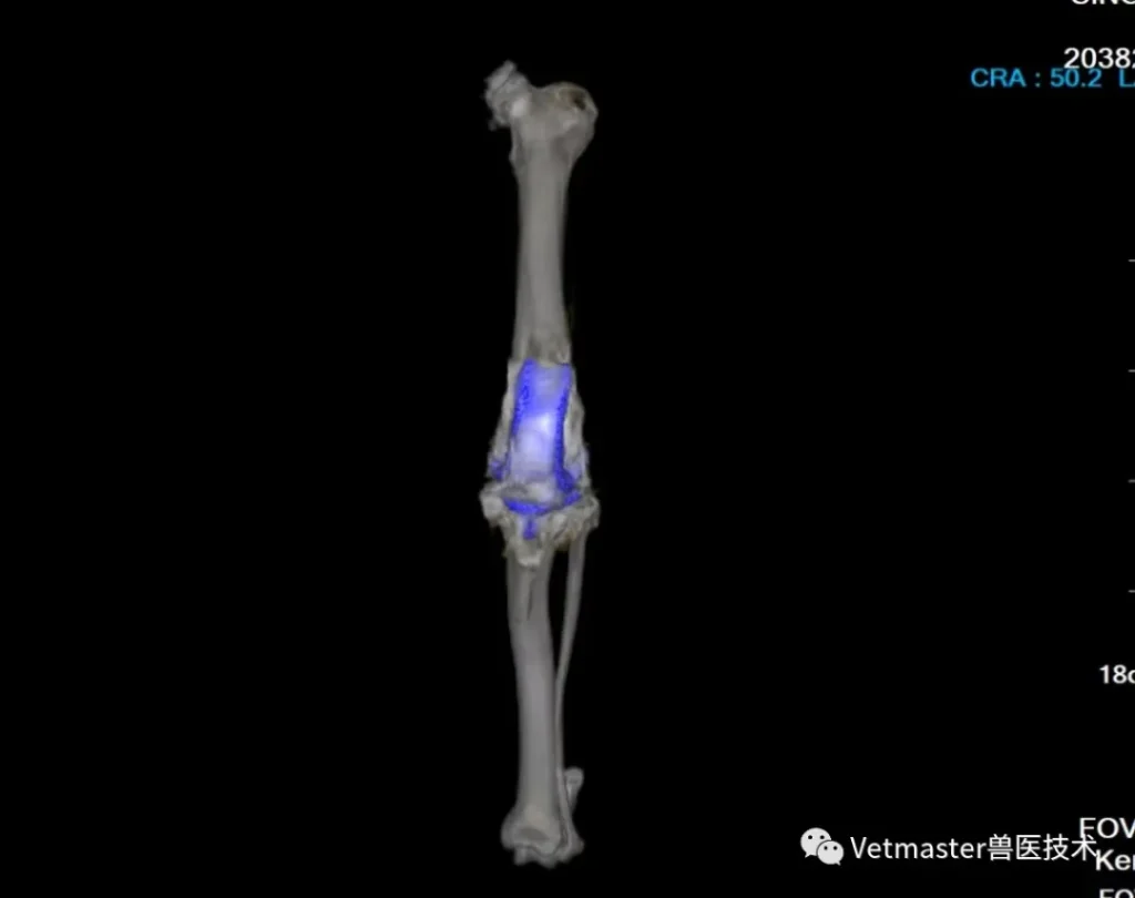
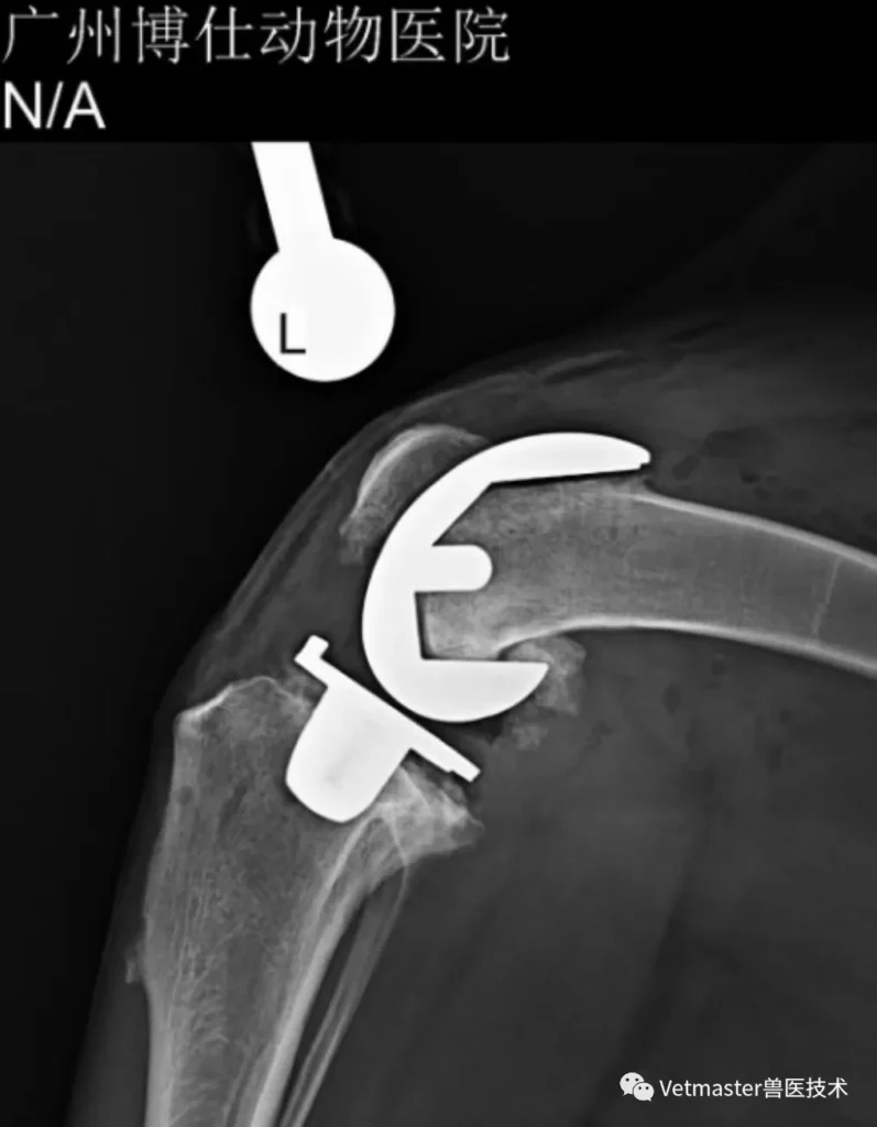
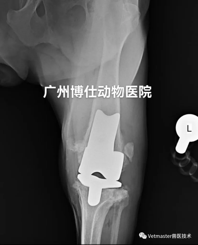
Intraoperative stability checks demonstrated full, unrestricted flexion and extension without any patellar maltracking or lateral toggle. The dog recovered smoothly from anesthesia under continuous multimodal pain management. Supported by our rehabilitation protocols, this powerful 41 kg athlete took his first comfortable, pain-free steps within days of surgery, embarking on a complete recovery that conventional medicine had declared impossible.
6. Clinical Reflections from the Front Lines
Canine total knee arthroplasty is not a surgery for the faint of heart:
- Master the Soft Tissues: In severe end-stage osteoarthritis, bone cuts are only half the battle. If you do not understand how to systematically release contractures while preserving collateral ligament integrity, you will never seat the implants properly.
- Refuse Compromises in Severe OA: When an animal presents with destroyed joint surfaces and torn cruciates, stop offering Band-Aid solutions. Realize when a joint is biologically dead, and step up to full joint replacement.
- Independent Technology Matters: Seeing a 41 kg Doberman walk out of our hospital on a biological knee system designed, engineered, and clinically proven right here in China is a testament to how far our veterinary orthopedic capabilities have come.
We don't need to look abroad anymore to save dogs from end-stage joint destruction.