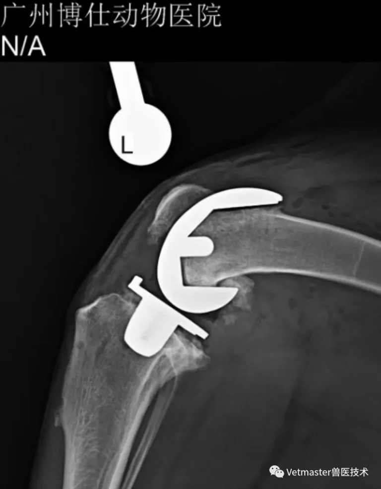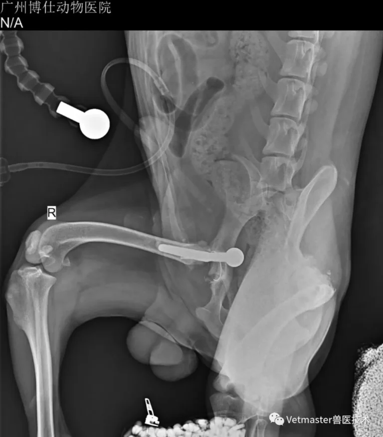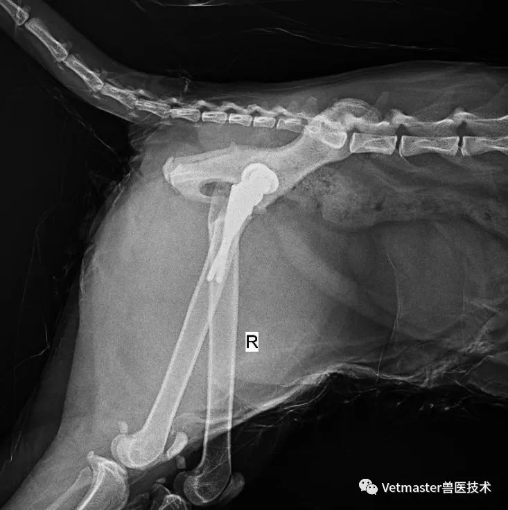
1. A Clinical Case That Demands a Closer Look
In veterinary joint reconstruction, the surgery does not conclude the moment the deep fascia is closed and the skin is sutured. Bone is not an inert scaffold; it is a dynamic, living tissue that constantly converses with the titanium implants we seat inside it.
Not long ago, a case at our clinic provided a textbook demonstration of this biological dialogue.
The patient was an 18 kg intact male Samoyed. Due to congenital hip dysplasia accompanied by severe joint laxity, I performed a left cementless total hip replacement (THR) using the Vetmaster biological dual-mobility system when he was 9 months old. Two months later, at 11 months of age, we completed the right THR.
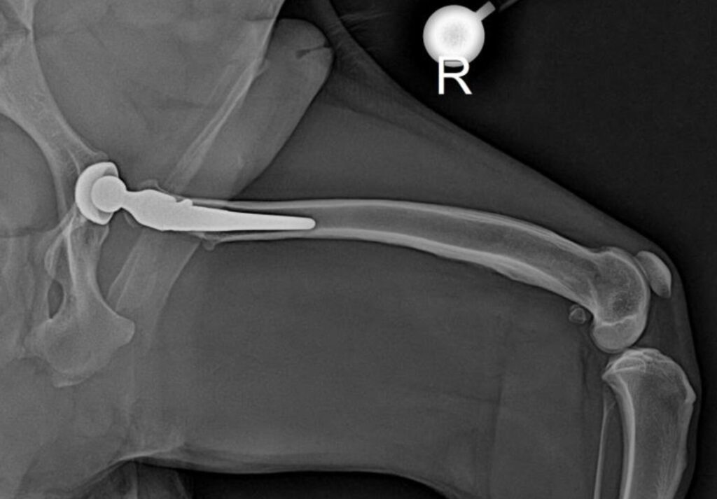
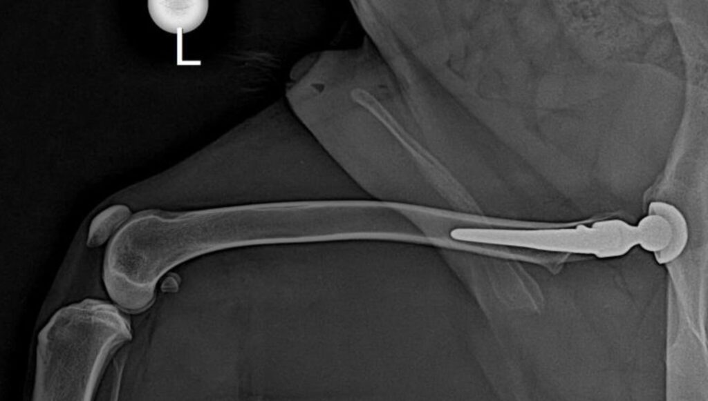
At his routine follow-up one month after the second surgery, orthogonal radiographs revealed a striking biological response: pronounced, smooth periosteal reaction along the distal cortex of the right femur, and mild, subtle periosteal thickening on the distal left femur.
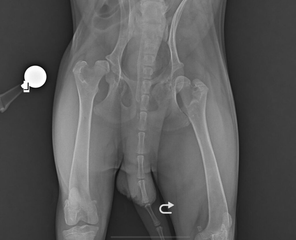
What exactly is a periosteal reaction? In basic histological terms, the periosteum—the vascular, osteoblast-rich "living sleeve" encasing the cortical bone—initiates accelerated bone formation in response to abnormal stimulation.
Following a cementless hip replacement, that "abnormal stimulation" is rarely an infection or malignancy. Far more often, it is the direct biological consequence of mechanical force redistribution.
2. Stress Shielding: The Mechanics of the Bypass
To understand why bone grows on the distal femur after a hip replacement, one must first grasp the core biomechanical concept of stress shielding.
Bone is an unapologetic "use-it-or-lose-it" organ. In the 19th century, the German anatomist Julius Wolff formulated Wolff’s Law: bone tissue continuously remodels and adapts its internal trabecular architecture and external cortical thickness in direct response to the mechanical loads it bears. Increase the load, and bone thickens; remove the load, and bone resorbs.
When we seat a biological prosthesis stem into the femoral canal, the natural load-transfer pathway of the hindlimb is altered:
- The structural stiffness of a medical-grade titanium alloy stem is significantly higher than that of the canine femur.
- As the dog bears weight, the axial load travels down the stiff prosthesis rather than traveling primarily through the proximal femoral cortex.
- The proximal bone is effectively "bypassed."
Because the proximal femoral cortex is no longer subjected to physiological load, Wolff’s law takes over: osteoclasts resorb unneeded bone mass, leading to proximal cortical thinning. This is classical proximal stress shielding.
However, mechanical load cannot simply vanish into thin air. The load that was bypassed proximally is transferred down the length of the stem and concentrated into the cortical bone surrounding the distal tip of the implant. The axial stress experienced by this distal cortex increases significantly. Sensing mechanical strain beyond its baseline threshold, the mechanosensitive periosteum initiates compensatory new bone formation—producing the smooth "periosteal reaction" we see on our screens.
This biomechanical chain is extensively validated in the literature:
- Experimental canine non-cemented THR models have demonstrated that high-stiffness stems generate approximately 26% proximal cortical stress shielding, whereas low-stiffness designs produce only about 7.5%.
- Long-term canine radiographic follow-ups consistently confirm this dual pattern: proximal bone resorption coupled with distal cortical hypertrophy.
Proximal resorption → distal load concentration → distal periosteal compensatory hyperplasia. The mechanical logic is completely self-consistent.
3. Why Did This Young Samoyed React So Vigorously?
While the stress-shielding mechanism applies to every hip replacement, the intensity of the periosteal response varies widely. In this young Samoyed, several patient-specific factors converged to amplify the biological reaction:
Factor 1: Active Growth Plates (9 to 12 Months of Age)
In medium-to-large canine breeds, the distal femoral physis typically bridges and closes between 10 and 12 months of age. As an Arctic sled dog, the Samoyed has a prolonged growth curve compared to smaller breeds. At 9 to 11 months, this dog's distal femoral physes and periosteal cambium layers were still in a hyper-metabolic growth phase. Juvenile bone remodeling responds far faster and far more aggressively to mechanical strain than mature adult bone.
A 2024 veterinary study specifically evaluated femoral dimensional changes after cementless THR in skeletally immature dogs (under 8.5 months of age). The data revealed that cortical width expansion on the operated femur was significantly greater than on the unoperated control—increasing by 4.6% at 10 mm distal to the greater trochanter and by 8.5% at the femoral mid-diaphysis. This cortical thickening represents active periosteal new bone formation.
Because this dog underwent sequential surgeries at 9 and 11 months, both femurs were subjected to the sudden mechanical shift of stress shielding while their growth plates were still biologically active.
Factor 2: Preoperative Severe Joint Laxity
Samoyeds are well-documented to carry a high genetic burden of hip dysplasia, with breed surveys reporting detection rates around 22.6%.
More importantly, this dog suffered from severe joint laxity before surgery. His proximal femurs had never experienced normal, concentric hip biomechanics. Comparative bone-mineral density studies in dogs with early osteoarthritis secondary to hip dysplasia reveal that chronic subluxation actually drives a 6% to 10% increase in subchondral and epiphyseal bone density.
However, increased density in the joint does not equal robust cortical bone quality in the diaphysis. It is a sign of reactive sclerosis under abnormal contact forces. The dog entered surgery with an abnormal, "subhealthy" proximal femoral cortex whose mechanical reserves were poorly prepared for the sudden axial stiffness of a cementless stem.
Factor 3: The Additive Effect
When you place a stiff titanium biological stem into a juvenile dog with hyperactive growth plates, whose proximal cortex was already compromised by chronic laxity, the stress-shielding cascade is magnified. Proximal bypass occurs more rapidly, and the load transferred distally hits the juvenile periosteum with greater relative force, easily pushing new bone formation past the visible radiographic threshold.
4. What This Means for Clinical Practice
For veterinary surgeons evaluating follow-up radiographs after cementless THR in young large-breed dogs, this case provides a vital clinical takeaway: distal femoral periosteal reaction is predominantly a mechanically driven adaptive remodeling process, not an automatic sign of disease or failure.
That said, recognizing a benign remodeling process does not mean dropping our clinical guard. A periosteal reaction is an objective signal that the local bone environment is undergoing intense mechanical reorganization. Our duty is to determine where that signal is heading:
- Benign Adaptive Remodeling (Monitor): If the periosteal reaction displays a smooth, solid, continuous margin, gradually stabilizes within 3 to 6 months post-op, merges into thickened cortex, and the patient remains completely comfortable and sound with no pain on deep palpation—this represents successful physiological adaptation. No intervention is required.
- Pathological Red Flags (Intervene): If the periosteal reaction continues to expand aggressively past 6 months, appears irregular, spiculated, or "moth-eaten," is accompanied by periprosthetic radiolucent lines, or coincides with recurrent lameness and pain—infectious periostitis or aseptic loosening must be treated as primary differentials.
Managing an infection on the porous, bone-ingrowth surface of a cementless biological stem is exponentially more challenging than dealing with a smooth cemented stem. When in doubt, serial radiographs, complete blood counts, and C-reactive protein (CRP) assays provide objective differentiation.
In orthopedic follow-up, never diagnose in isolation based on a single radiographic shadow. Evaluate the implant, the mechanical load axes, and most importantly, the living dog walking in front of you.