1. The Dangerous Myth of "Conservative Management"
In small animal practice, patellar luxation is frequently trivialized. Far too many owners—and unfortunately, some veterinarians—believe that as long as a dog is not screaming in pain, a luxating kneecap can simply be "managed" with joint supplements, painkillers, and restricted activity.
What they fail to realize is that patellar luxation is an unforgiving mechanical conflict. Every time that patella hops over the trochlear ridge and snaps back into place, it acts like coarse sandpaper across the hyaline cartilage. Medication can suppress temporary inflammation, but it cannot stop bone from grinding away bone.
This 4-year-old intact male Pomeranian weighing 5.5 kg was a direct casualty of that delay. He had been diagnosed with patellar luxation at an outside hospital more than a year earlier. His owners opted for conservative therapy, hoping the condition would magically stabilize. Instead, his left hindlimb lameness progressively worsened over the preceding months until he was persistently limping and reluctant to bear weight.
When they brought him to Guangzhou Boss Animal Hospital, the examination room told the whole story:
- Palpation confirmed a Grade 3 medial patellar luxation (MPL) in the left stifle. The patella was luxated most of the time, although it could be manually reduced into the groove with the limb extended.
- The moment I gently flexed and extended the joint, a coarse, unmistakable articular crepitus vibrated through my fingertips.
- The joint was bone-dry, stiff, and distinctly painful on full extension.
2. The Radiographic Crossroads: Why Traditional Deepening Fails
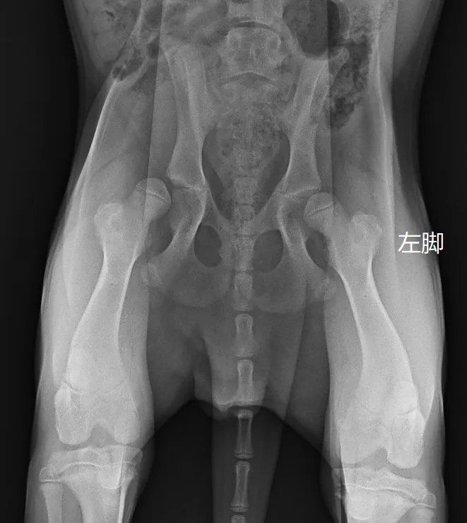
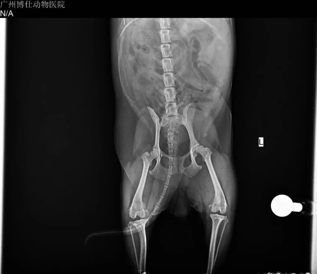
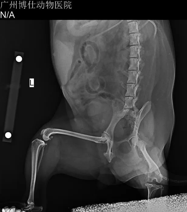
Orthogonal stifle radiographs confirmed the mechanical collapse of the patellofemoral joint:
- The left patella was displaced medially.
- The medial trochlear ridge was severely worn, flattened, and surrounded by dense subchondral sclerosis and extensive periarticular osteophytes.
- The natural trochlear architecture was effectively erased.
At this stage, a veterinary surgeon faces a defining decision:
- Option A: Traditional Sulcoplasty (Wedge/Block Recession) + Tibial Tuberosity Transposition (TTT):
This is what ninety percent of clinics would attempt. You cut a wedge of bone, deepen the sulcus, and shift the crest. But in a joint with Grade 3 chronic disease, the cartilage is already gone. Carving a deeper trench out of denuded, sclerotic bone simply forces the patella to articulate against an unyielding, rough bony bed. The patella might stay in the center, but the friction remains. The dog will continue to suffer from chronic, low-grade osteoarthritic pain and a permanent hitch in its gait. - Option B: Patellar Groove Replacement (PGR):
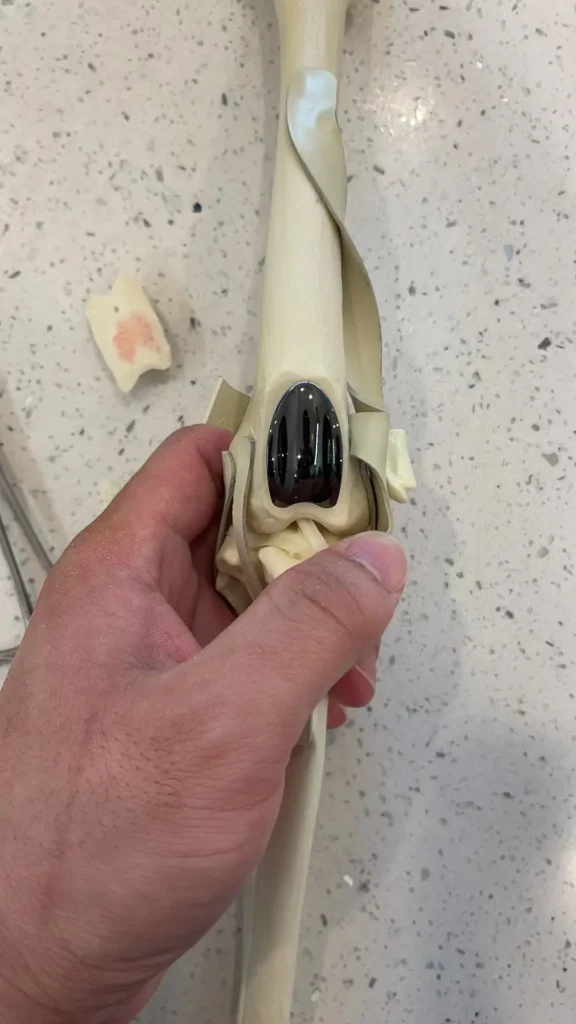
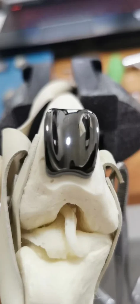
Instead of scraping raw bone, we resect the worn trochlear surface and anchor an ultra-smooth, biocompatible metal prosthesis engineered specifically to restore a frictionless tracking channel.
I have been performing KYON PGR surgeries since back in 2013—one of the first surgeons to introduce the technique in mainland China—and my clinic maintains a comprehensive inventory of Swiss KYON implants across all sizes. After discussing the realities with the owner, they chose to go with the definitive solution: a Swiss-manufactured KYON PGR.
3. Surgical Execution: Swiss KYON Size 2 PGR
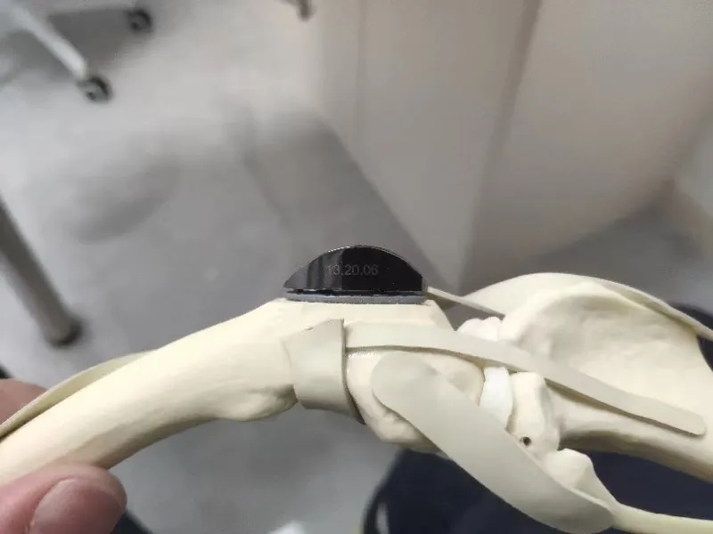
Following precise radiographic template measurements, I selected a Size 2 KYON PGR prosthesis.
The surgical procedure requires strict precision and zero margin for error:
- Arthrotomy and Inspection:
Entering the joint capsule revealed exactly what the crepitus had warned us about. The articular cartilage of the trochlea was completely destroyed, exposing dull, ivory-like eburnated bone flanked by massive osteophytic ridges. - Reconstruction of the Femoral Bed:
Using dedicated KYON precision milling jigs, I resected the diseased trochlear surface down to healthy, vascularized cancellous bone, ensuring the cut perfectly aligned with the anatomical coronal and sagittal axes of the distal femur. - Implantation & Fixation:
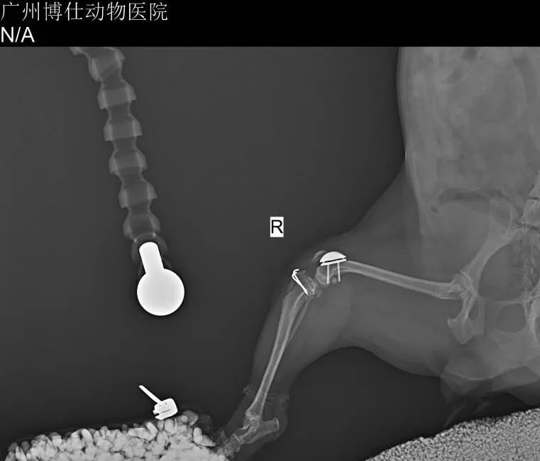
The Size 2 titanium baseplate and low-friction, diamond-like coated prosthetic groove were secured using monocortical titanium locking screws. The prosthesis achieved seamless, rock-solid contact with the host bone. - Tracking & Alignment Check:
Once the PGR was anchored, we reduced the patella. It engaged the artificial groove instantly. Throughout the full range of physiological flexion and extension, the patella glided with silky, fluid smoothness—zero catching, zero crepitus, and zero tendency to subluxate medially.
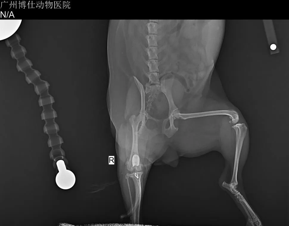
Postoperative orthogonal radiographs confirmed textbook positioning: the PGR prosthesis sat flush within the anatomical sulcus, restoring normal depth and lateral support, while the patella centered cleanly within the groove.
4. Clinical Reflections: A Surgeon’s Golden Rule
This case highlights core truths about feline and canine knee pathology that I emphasize repeatedly in my lectures across the country:
- Cartilage Is Irreplaceable:
Once articular cartilage is ground down, biological healing cannot bring it back. Fibrocartilage repair is mechanically inferior and wears out rapidly. That is why I tell every owner: Grade 2 or higher patellar luxation should be operated on early, before the cartilage wears through. Waiting until the joint creaks like an un-oiled hinge only forces you into expensive salvage procedures. - The Truth About Prostheses vs. Nature:
I often share this clinical philosophy with colleagues:"Native, living cartilage is always superior to any metal replacement; but an engineered metal prosthesis is infinitely better than bone grinding painfully against bare bone."
A metal groove cannot replicate living biology, but for an animal whose native joint has been worn down to raw bone, PGR is a miracle of mechanical engineering that erases pain overnight. - Recognizing Stifle Osteoarthritis:
Trochlear groove osteoarthritis secondary to chronic luxation is one of the most under-diagnosed and under-treated conditions in small dogs. Surgeons often fix the alignment and wonder why the dog still limps six months later. If the cartilage is gone, realigning the patella is only half the job—you must replace the friction surface.
Watching this little Pomeranian put his foot down cleanly and comfortably post-op was a reminder of why precision orthopedics matters: when you eliminate joint friction, you give an animal back its joy of life.