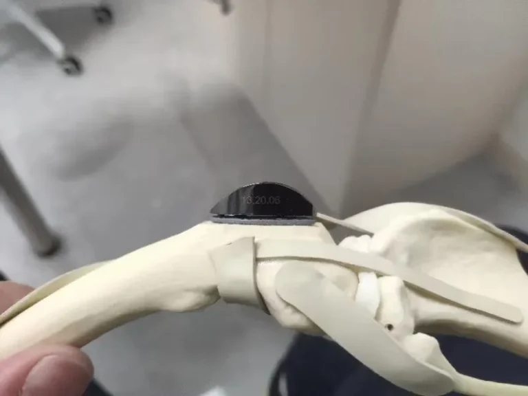
1. The Surgeon's Priority: Joint Salvage vs. Joint Preservation
A lot of veterinarians and pet owners know me primarily for total hip replacements (THR). Having performed hundreds upon hundreds of hip replacements, I understand the life-changing power of an artificial joint. But make no mistake: THR is a salvage procedure. You replace a joint because the native cartilage has been ground away, the acetabular rim is eburnated, and the joint is past the point of biological return.
Given the choice, I would far rather help an animal keep its own natural, healthy hip.
If we detect canine hip dysplasia (CHD) early—before joint incongruity grinds down the articular cartilage and sets off irreversible degenerative joint disease—we don't need to replace the hip later in life. We can structurally correct it. That is the fundamental clinical philosophy behind juvenile hip screening and Double Pelvic Osteotomy (DPO).
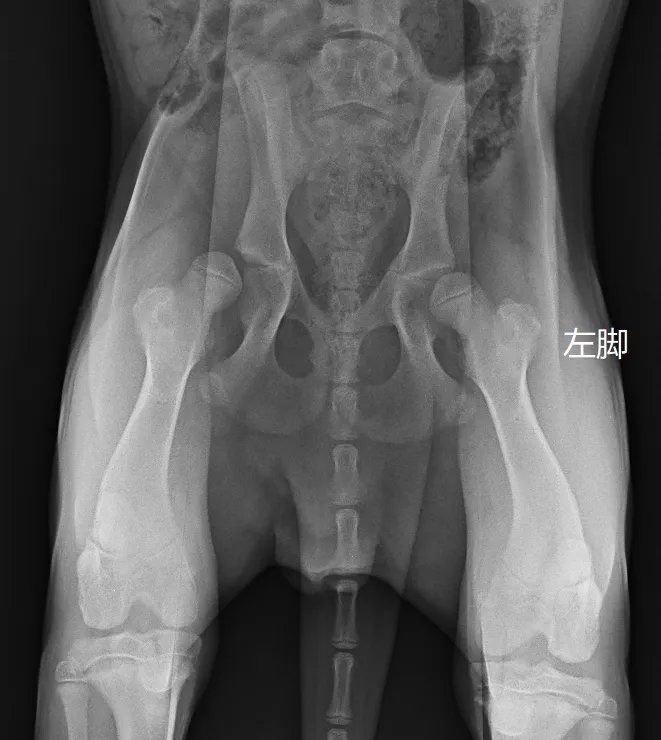
When this 6-month-and-24-day-old intact male Golden Retriever weighing 23 kg was brought to Guangzhou Boss Animal Hospital, his owner had noticed a distinctly abnormal, loose hindlimb gait. The clock was ticking. In juvenile large-breed dogs, the therapeutic window for joint-preserving osteotomies closes rapidly between 5 and 8 months of age.
2. Quantitative Screening: The Science of Choosing the Right Side
You cannot evaluate canine hip dysplasia on a juvenile dog with a quick conscious physical exam or a single standard radiograph. As I have demonstrated repeatedly in my teaching and publications, juvenile hip evaluation requires a structured, multi-step protocol under deep sedation: assessing the Ortolani sign, measuring the specific angles of reduction and subluxation, and capturing orthogonal views alongside PennHIP distraction radiographs.
Our quantitative evaluation of this young Golden Retriever revealed contrasting pathologies:
- Left Hip:
- Ortolani sign: Positive.
- Angle of Subluxation (AS): 18°.
- Angle of Reduction (AR): 37°.
- Distraction Index (DI): 0.71.
- Articular cartilage: Preserved, with no radiographic signs of secondary osteophytosis or dorsal acetabular rim erosion.
- Right Hip:
- Ortolani sign: Positive.
- Angle of Subluxation (AS): 21°.
- Angle of Reduction (AR): 38°.
- Distraction Index (DI): 1.03.
These numbers dictated our surgical boundary. In juvenile hip preservation, a Distraction Index above 0.85 is an established contraindication for DPO. When joint laxity is that extreme (as in the right hip with DI 1.03), rotating the acetabular segment by 25° or 30° cannot provide sufficient mechanical capture of the femoral head; forcing an osteotomy in such a joint inevitably leads to persistent subluxation, acetabular impingement, and early failure. The right hip was not a candidate for osteotomy; its long-term future will be monitored and managed, likely culminating in a total hip replacement once skeletal maturity is reached.
The left hip, however, sat right in the sweet spot. With a DI of 0.71, favorable reduction mechanics, and intact cartilage, it was an ideal candidate for Double Pelvic Osteotomy (DPO).
3. The Procedure: Double Pelvic Osteotomy (DPO)
I learned the nuances of early hip screening and pelvic osteotomy directly from my mentor, Dr. Aldo Vezzoni, in Cremona, Italy. Dr. Vezzoni pioneered DPO as an evolution of the older Triple Pelvic Osteotomy (TPO).
Unlike TPO, which requires cutting the ilium, pubis, and ischium, DPO involves osteotomies of only the ilium and pubis, leaving the ischial table structurally intact. This preservation of the ischial arch maintains the intrinsic rigidity of the pelvic canal, dramatically reduces postoperative implant stress, prevents pelvic collapse, and allows for immediate, comfortable bilateral weight-bearing without external coaptation.
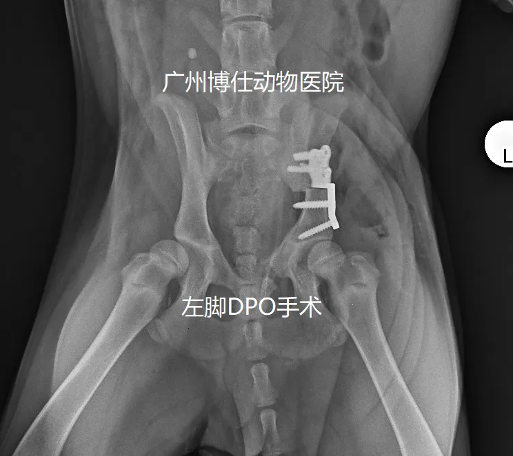
Under strict sterile conditions, I performed the left-sided DPO:
- A pubic ostectomy was executed to release the ventral constraint on the hemipelvis.
- An iliac osteotomy was performed perpendicular to the dorsal pelvic axis.
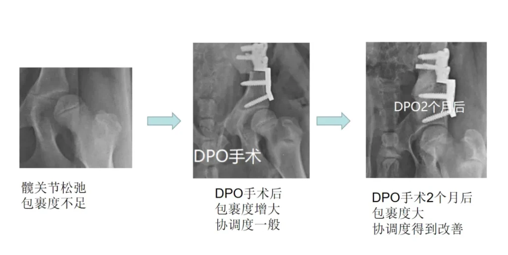
- Using a specialized, pre-angled DPO plate, I rolled the acetabular segment ventrolaterally, effectively rotating the socket downward over the top of the femoral head.
Immediate postoperative radiographs confirmed that the left femoral head was now deeply seated beneath the rotated dorsal acetabular rim. The mechanical capture was immediate and rigid.
4. Two-Month Follow-Up: Evidence in the Muscle
While immediate post-op films show mechanical coverage, the real biological proof of DPO reveals itself over the subsequent weeks of growth.
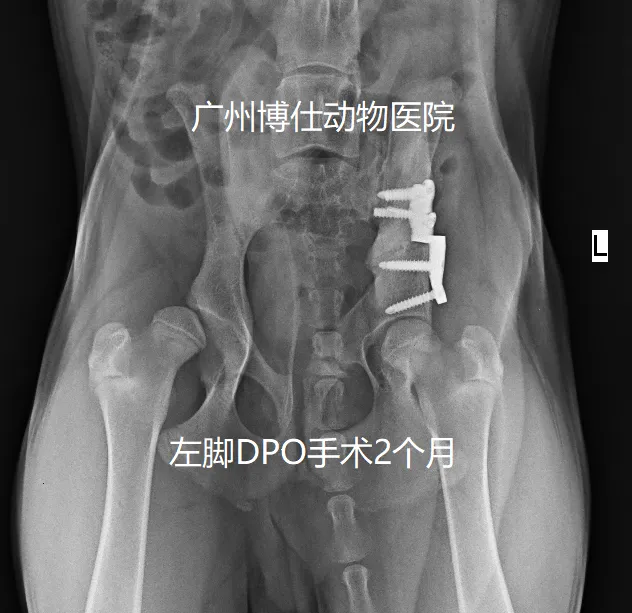
At his two-month re-examination:
- Radiographic Congruity: The serial radiographs demonstrated exceptional integration. As the puppy grew over the preceding eight weeks, the femoral head remodeled smoothly within the newly deepened acetabulum. The joint congruity had markedly increased, and there was zero progression of secondary osteoarthritis.
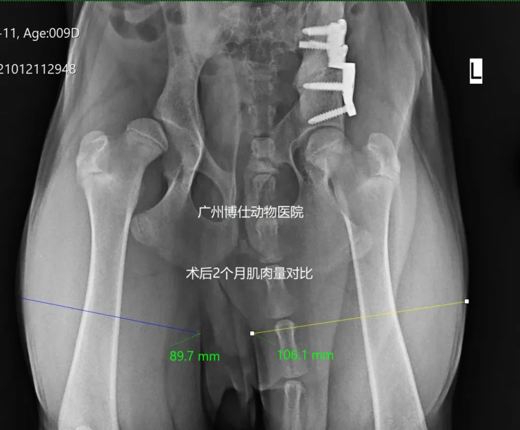
- Objective Muscle Mass (The 20% Metric):
When we measured the dog's thigh circumference, the muscle diameter of the operated left hindlimb was roughly 20% larger than that of the unoperated right hindlimb.
In orthopedic follow-ups, muscle mass doesn't lie. Because the painful stretching and instability of the left joint capsule had been neutralized by the DPO, the dog had naturally and preferentially loaded his left leg for two months. Disuse muscle atrophy had completely reversed on the surgical side, providing objective, indisputable evidence of restored comfort and joint function.
5. Clinical Reflections: Catch Them Early, Save the Joint
This Golden Retriever’s journey underscores why I continue to advocate tirelessly for early orthopedic screening in China:
- Screen Early (4.5 to 8 Months):
Waiting until a dog is limping heavily at two years old is waiting too long. By that time, the cartilage has worn down, osteophytes have populated the joint, and the window for DPO is closed forever. Puppies of high-risk breeds (Golden Retrievers, Labradors, German Shepherds, Corgis) should be screened under sedation between 4.5 and 6 months of age. - Preservation Over Salvage:
A well-executed DPO costs less, carries far lower catastrophic risks than joint arthroplasty, and allows the animal to live out its entire life on its own living bone and cartilage. - Know Your Limits:
The success of DPO lies as much in knowing when not to cut as in knowing how to cut. Attempting DPO on the right hip (DI 1.03) would have been an exercise in surgical arrogance that harmed the dog. By respecting the biological limits (DI < 0.85), we protected one hip for life while keeping our options open for the other.
Every time I see an operated DPO puppy run across a field on two native, pain-free hips, it reaffirms my conviction: early diagnosis is the highest form of veterinary medicine.