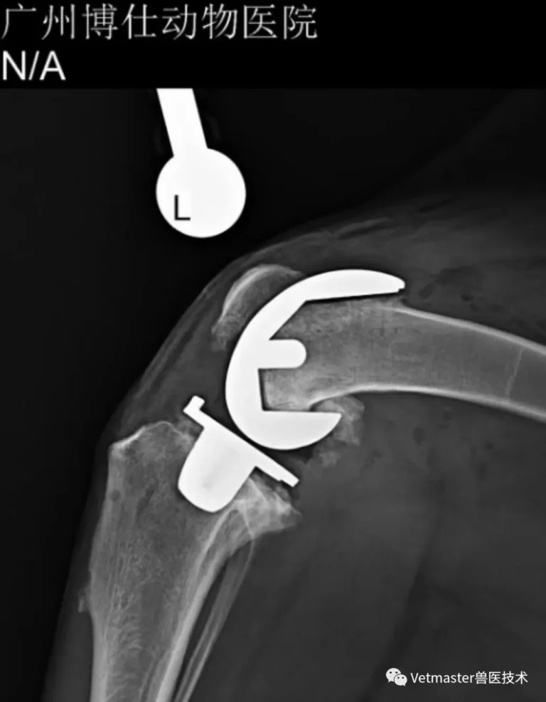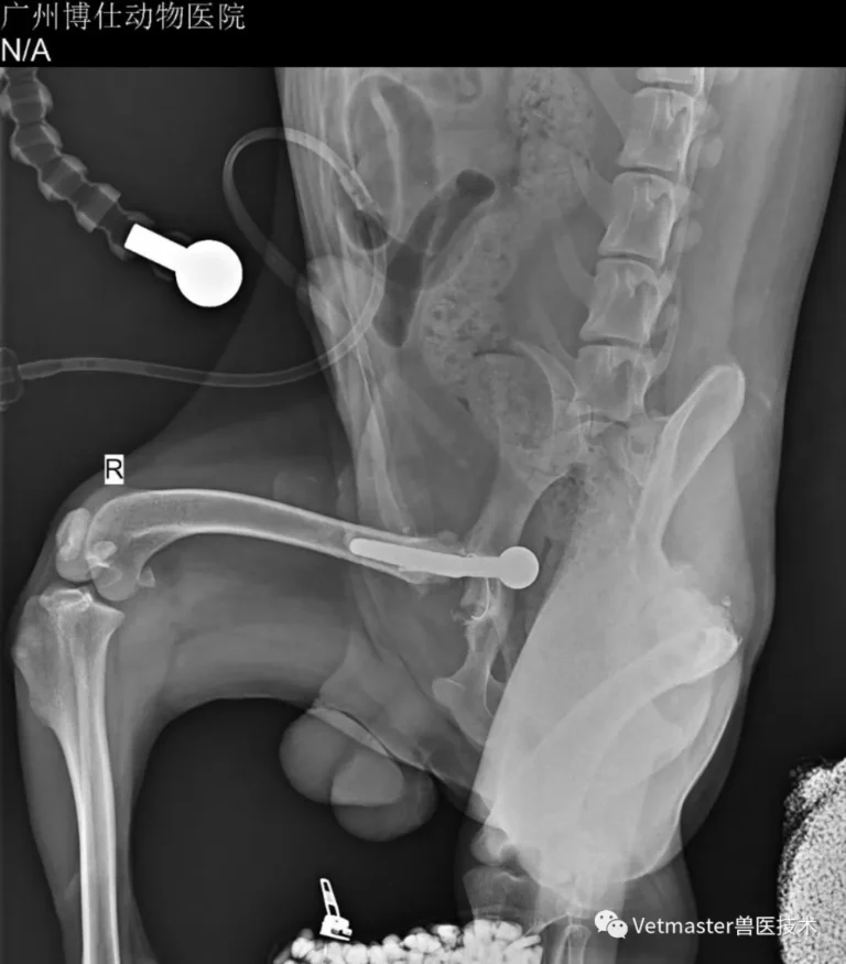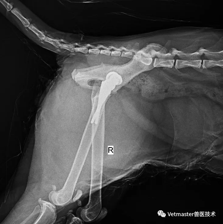
1. The "Corgi Factor": Why This Breed Keeps Surgeons on Their Toes
Corgis are undeniably charming, but from an orthopedic surgeon's perspective, they are a bundle of structural contradictions.
They are an orthopedic high-risk breed across the board:
- Highly predisposed to medial and lateral patellar luxation;
- Prone to cranial cruciate ligament (CCL) rupture;
- Highly susceptible to intervertebral disc disease (IVDD) leading to sudden paralysis due to their disproportionately long lumbar spines;
- Genetically prone to hip dysplasia and severe hip laxity;
- And to compound matters, they have an innate tendency toward obesity, which exponentially multiplies the mechanical stress on their already vulnerable joints.
Because Corgis are heavily muscled, they have a remarkable capacity to mask joint pain in the early stages. Instead of visibly limping, they use raw gluteal muscle tension to fight against hip joint laxity, shifting their weight forward and undulating their lower back to propel themselves forward.
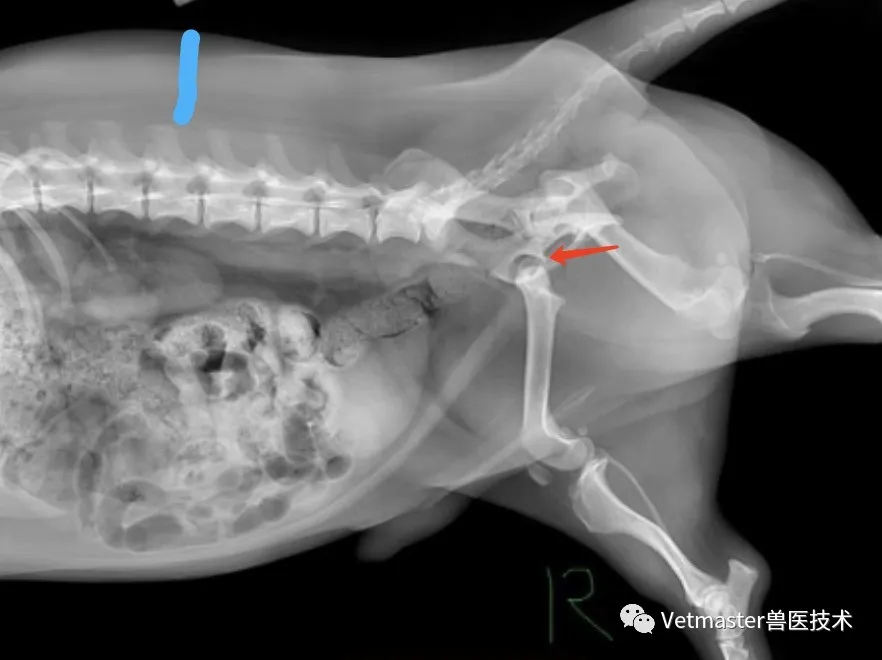
That was the exact presentation of this patient: an intact male Corgi, just 1 year old, weighing a solid 14 kg. His owner brought him to Guangzhou Boss Animal Hospital after noticing that his hips and waist swayed aggressively from side to side whenever he walked.
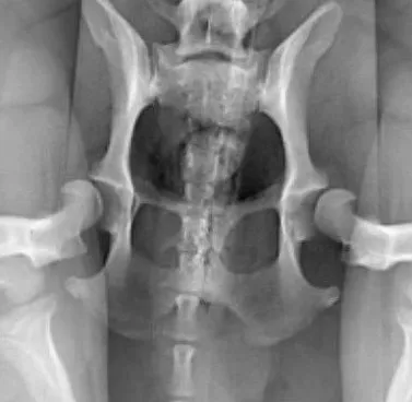
Physical palpation under sedation and orthogonal radiographs confirmed severe bilateral hip laxity. Fortunately, because he was young, the articular cartilage had not yet suffered massive arthritic flattening or eburnation. But with laxity this severe, waiting for arthritis to destroy the joint was pointless. We decided to proceed with staged bilateral Total Hip Replacement (THR) using the Vetmaster cementless dual-mobility system.
2. Stage One: Left Total Hip Replacement
I addressed the left hip first.
Operating on a Corgi is never a standard, textbook procedure:
- The Soft-Tissue Corridor: Corgis have exceptionally short femurs buried beneath thick layers of subcutaneous fat and dense gluteal musculature. The surgical field is deep, narrow, and easily obscured, requiring meticulous soft-tissue elevation and constant vigilance against pelvic tilting.
- Medullary Proportions: The feline-like wide medullary canal relative to their cortical diameter demands precise rasping to achieve optimal canal fill without splitting the proximal cortices.
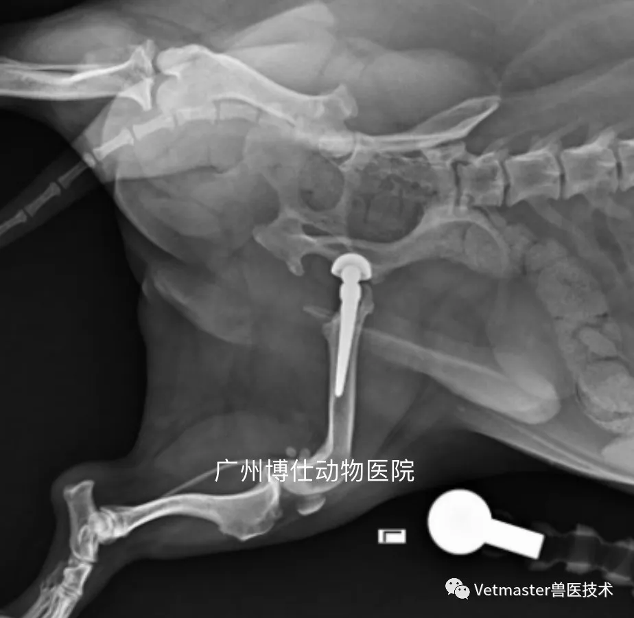
Working carefully through our standard lateral approach, I reamed the acetabulum down to healthy subchondral bone, preserving the dorsal rim, and placed a 16 mm Vetmaster biological cup. The femoral canal was broached to receive a biological stem, achieving an immediate, rock-solid press-fit occupying over 85% of the canal.
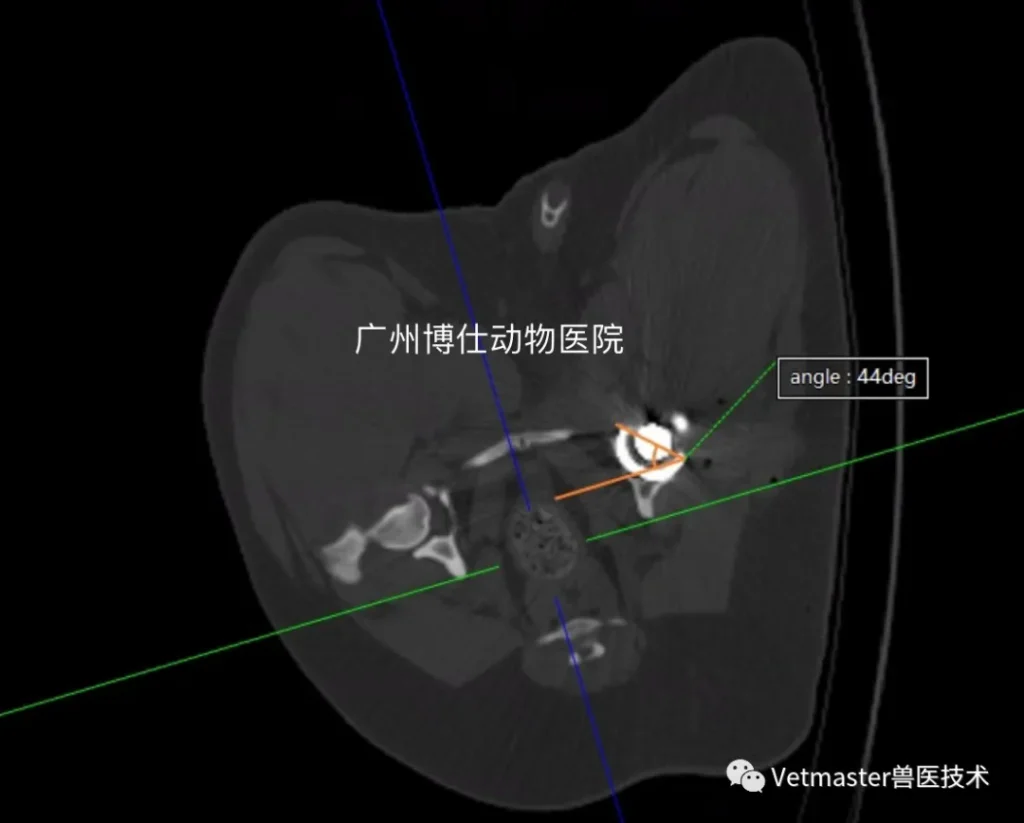
Postoperative multi-planar CT evaluation confirmed excellent positioning:
- Cup Abduction Angle: Measured at 44°, sitting right in the ideal 45° physiological target.
- Cup Version: Perfectly matched the natural physiological retroversion angle.
- Stem Alignment: Centralization was spot-on, with excellent axial and rotational stability.
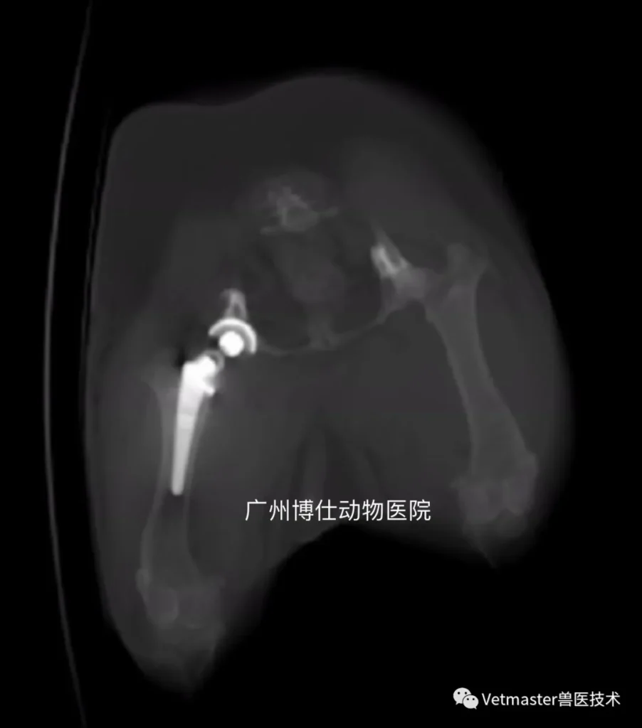
The dog recovered rapidly. At his one-month follow-up, the left limb was bearing full, fluent weight, and the surgical site was pristine. With the left hip fully stabilized, the owner requested that we move forward with the contralateral side.
3. Stage Two: Right THR and Achieving Bilateral Symmetry
One month after the first surgery, we brought the Corgi back into our clean-room surgical suite for the right Total Hip Replacement.
With the left hip now providing robust, pain-free weight-bearing, the right-sided procedure went with absolute fluidity:
- The acetabular bed was prepared manually, and a second biological dual-mobility cup was impacted into place.
- The right femoral canal was prepared, and the matching biological stem was seated cleanly into the diaphyseal axis.
- We reduced the dual-mobility ceramic head within its mobile polyethylene liner, verifying full, unrestricted range of motion.
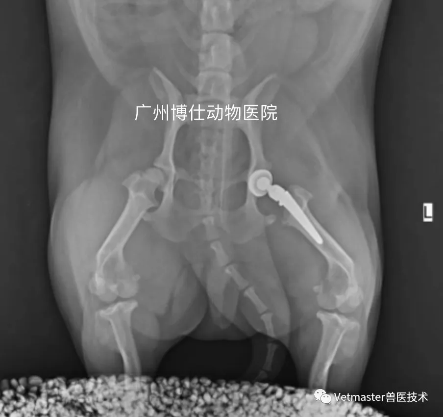
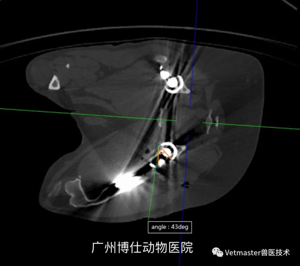
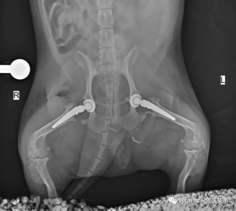
Postoperative orthogonal radiographs and 3D CT volumetric reconstructions demonstrated textbook bilateral symmetry:
- The right acetabular cup achieved an abduction angle of 43°—virtually identical to the left cup's 44°.
- Both femoral stems sat at identical depths and physiological anteversion angles within their respective canals.
4. Clinical Reflections on Corgi Hip Arthroplasty
Managing hip replacements in chondrodystrophic, heavily muscled breeds like Corgis yields vital clinical lessons:
- Never Underestimate the "Wiggle": An exaggerated waist-swaying gait in a young Corgi is almost never a cute behavioral quirk; it is a textbook compensatory adaptation to severe hip laxity. Catching and replacing these hips before irreversible secondary arthritis destroys the acetabular bone stock makes for a vastly smoother surgical outcome.
- Respect the Adipose and Muscular Anatomy: In short-legged, muscular dogs, you cannot rush your surgical approach. Take the time to clear the dense subcutaneous fat and achieve pristine visualization of the dorsal acetabular rim.
- Dual-Mobility Provides the Safety Margin: Corgis have wide pelvic angles and explosive muscular drive. Utilizing a dual-mobility ceramic articulation gives these active little dogs the extra jump distance needed to eliminate post-op luxation risks during rapid recovery.
Today, this 14 kg Corgi has two symmetrical, biological artificial hips that will support his active life for the next decade and a half—completely free from joint pain and waist-swaying instability.