1. The Clinical Dilemma Behind Canine Knee Replacements
Back when I performed this surgery, canine total knee arthroplasty (TKA) was practically uncharted territory in China. In human orthopedic surgery, total knee replacement is routine—hundreds of thousands of procedures are done every year with decade-long success rates exceeding 90%. But in small animals, particularly in our domestic market, cases were virtually nonexistent.
The barrier wasn't just surgical technique; it was implants and instrumentation. The proprietary systems were monopolized abroad, extraordinarily expensive, and simply unavailable off the shelf when you encountered a patient who genuinely needed one. When a dog develops irreversible end-stage knee osteoarthritis—especially when long-standing instability has ground down the articular cartilage and destroyed the meniscus—standard osteotomies like TPLO or TTA come too late to fix the pain. You are either forced into lifelong NSAID compromises, or you step up and rebuild the joint from scratch.
Faced with a joint that conventional methods could no longer salvage, I decided to take a bold route: designing a patient-matched, biological total knee replacement using custom 3D metal printing.
2. Case Presentation: A Chronic, Pain-Racked Stifle
The patient was a 25 kg Husky referred to us with persistent, marked left hindlimb lameness that had severely compromised his everyday life.
On physical examination:
- Obvious joint effusion and coarse crepitus were palpable throughout both flexion and extension of the left stifle.
- The tibial compression test was positive.
- The cranial drawer test was ambiguous (±), typical of chronic cranial cruciate ligament (CCL) tears where extensive periarticular fibrosis and osteophyte formation partially mask joint laxity.
- Severe joint pain was elicited upon manipulation.
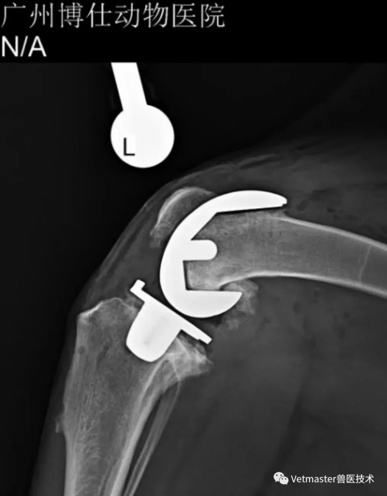
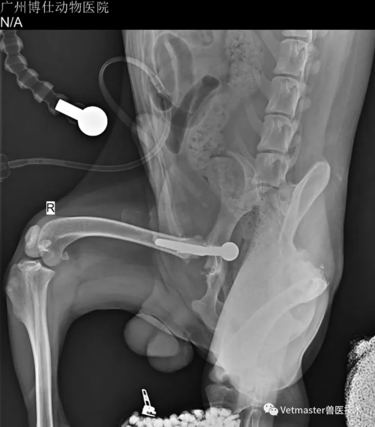
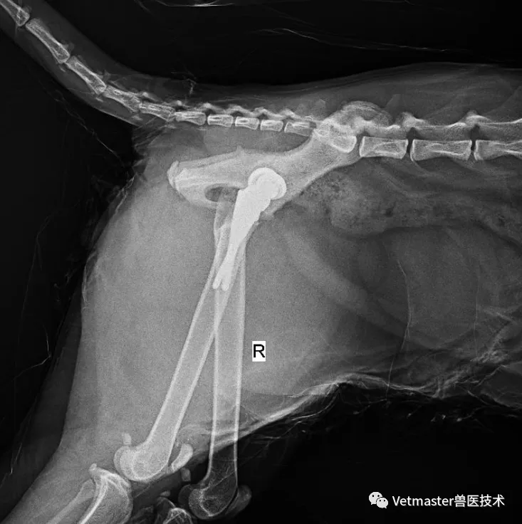
Orthogonal radiographs and computed tomography (CT) confirmed what our hands already suspected: severe, end-stage degenerative joint disease with ragged joint margins, dense periarticular osteophytosis, and structural disruption of the medial compartment and meniscus. A thorough orthopedic and radiographic screening of both hips revealed no significant abnormalities, confirming that his disability stemmed entirely from this destroyed knee joint.
3. Engineering a Solution: From CT Data to 3D Metal Printing
Because commercially viable, standardized biological knee implants of appropriate scale did not exist domestically at the time, I initiated a collaborative effort with our engineering counterparts. Guangzhou Vetmaster teamed up with Associate Professor Zhang Jun and the doctoral materials science team at the Institute of New Materials, Guangdong Academy of Sciences.

Together, we formed an interdisciplinary medical-engineering team to tackle this from the ground up:
3.1 Component Design & Biomaterials
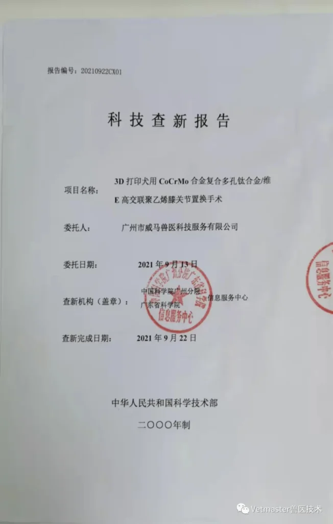
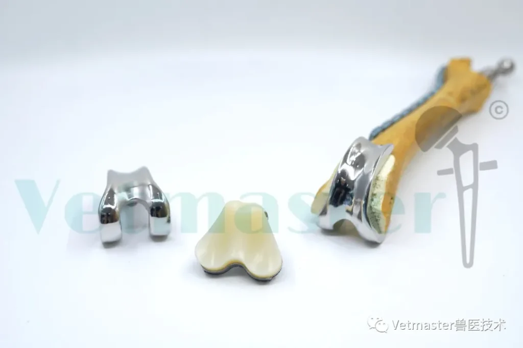
Using the dog's high-resolution CT volumetric data, we reconstructed the bone geometry and designed a three-part biological total knee system: a femoral condylar component, a tibial plateau tray, and a meniscal bearing insert. The bearing interface incorporated Vitamin E highly cross-linked polyethylene (VE-UHMWPE), matched against a CoCrMo alloy articulated surface, with porous titanium structures engineered directly onto the bone-contact surfaces to ensure bone ingrowth. (This specific material configuration was later confirmed by the Ministry of Science and Technology's official novelty search report as a world-first in canine TKA).
3.2 Toxicology and Mechanical Validation
Prior to clinical application, the printed alloy implants underwent rigorous mechanical stress testing and biocompatibility assessments to verify fatigue strength and implant-grade safety standards.
3.3 Custom Disposable Instrumentation
A major pitfall in custom joint surgery is the absence of dedicated cutting jigs. We used 3D printing to produce a complete set of patient-matched, single-use surgical cutting guides and trial components.
3.4 Benchtop Simulation on 1:1 Models
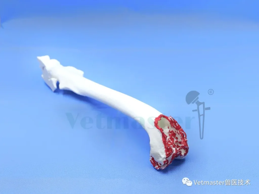
I never go into an unprecedented procedure blind. Before sterilizing the final implants, we 3D-printed a 1:1 replica of the patient's femur and tibia in plastic. In our prep room, I performed full step-by-step trial runs on the model—osteotomies, alignment verification, trial reductions, and range-of-motion assessments. This dry run ironed out our cut angles and confirmed that our instrumentation seated flush against the cortical contours, eliminating unwelcome surprises in the real operating theater.
4 Surgical Outcome and Follow-up
My surgical team at Guangzhou Boss Animal Hospital performed the implantation.
Thanks to our thorough benchtop rehearsals, the bone cuts and implant placement proceeded smoothly. The porous surfaces achieved an immediate, rock-solid press-fit against the prepared cancellous beds of the distal femur and proximal tibia. Postoperative radiographs confirmed textbook implant positioning, physiologic alignment, and an anatomical fit with the host bone.
The immediate postoperative recovery was remarkable:
- Day 1 Post-Op: The dog was already weight-bearing comfortably on the operated limb, enjoying a gentle, pain-free walk—a dramatic contrast to his preoperative guarded posture.
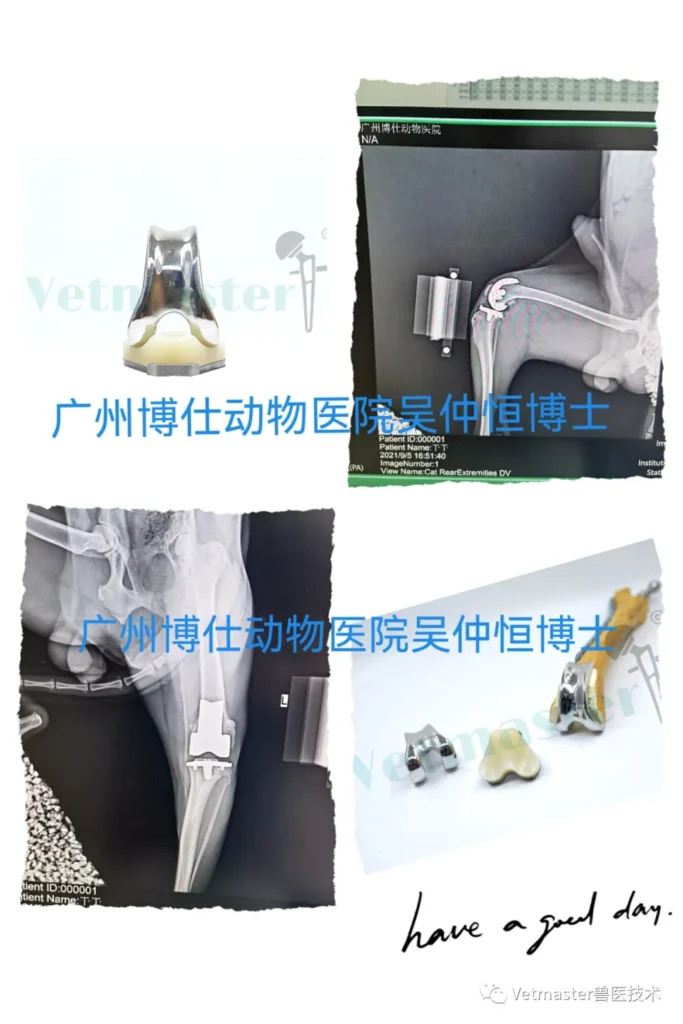
- 2.5 Months Post-Op: The owner shared video footage from an outdoor gathering. The Husky was galloping and playing across open grass without restraint. Looking at his fluent stride, it was nearly impossible to tell he had undergone a major joint reconstruction just ten weeks prior.
5. Clinical Reflections from the Operating Table
Every custom procedure comes with zero margin for error. When you venture where there are no existing textbooks or standardized kits, success demands three things: an intimate grasp of functional anatomy, a watertight preoperative plan, and genuine engineering collaboration.
From my clinical experience across hundreds of joint replacements, a few pragmatic takeaways stand out:
- TKA vs. THR Dynamics: Compared to total hip replacement, small animal total knee arthroplasty features an inherently more constrained, bicondylar architecture. When designed and seated properly, post-op stability is exceptional and mechanical luxation is considerably less common than in hips. However, stifle rehabilitation requires patience; soft tissue adaptation around the knee takes time, and overall functional remodeling spans four to six months.
- Salvaging the "Unsalvageable" Stifle: When chronic CCL rupture or chronic patellar luxation has chewed through the joint cartilage down to eburnated bone, osteotomies like TPLO cannot reverse the arthritis. In those irreversible, end-stage scenarios, biological TKA is not an aggressive luxury—it is the only definitive cure that relieves pain by replacing the worn-out friction surfaces.
This case was a milestone for my team, demonstrating that with disciplined surgical planning and home-grown 3D printing technology, we can solve cases once deemed untreatable in Chinese veterinary practice.