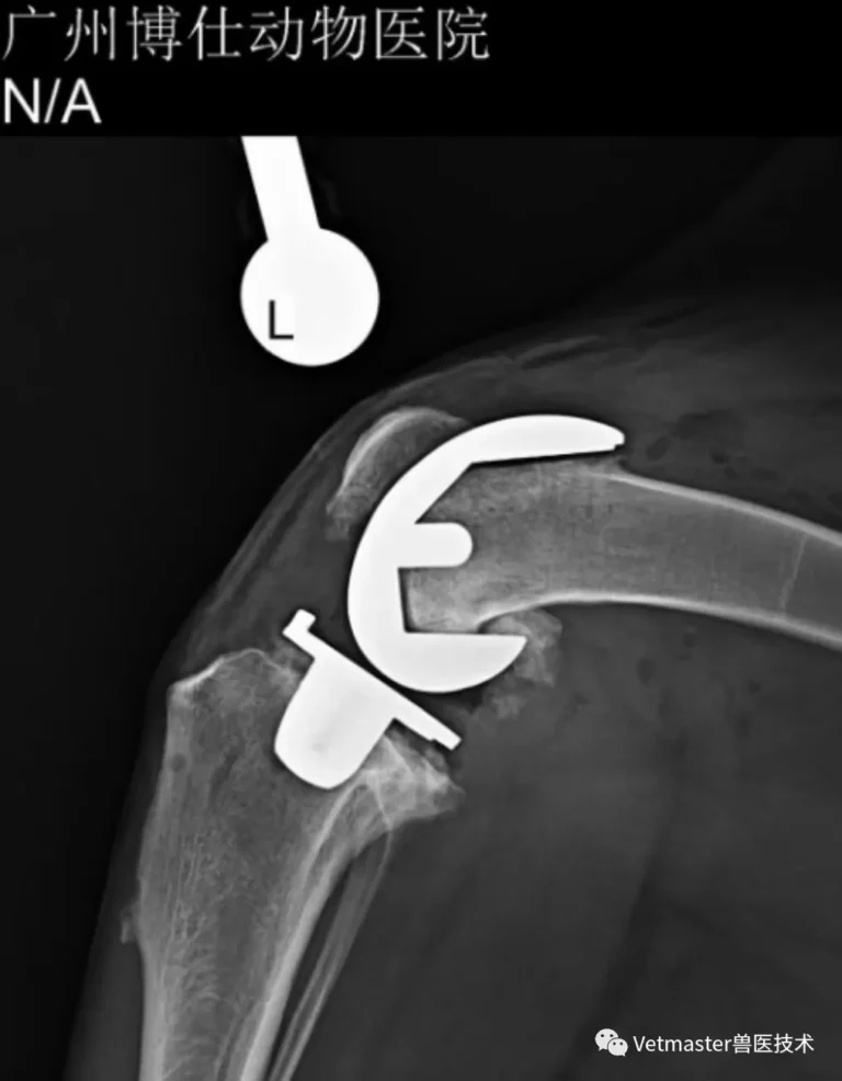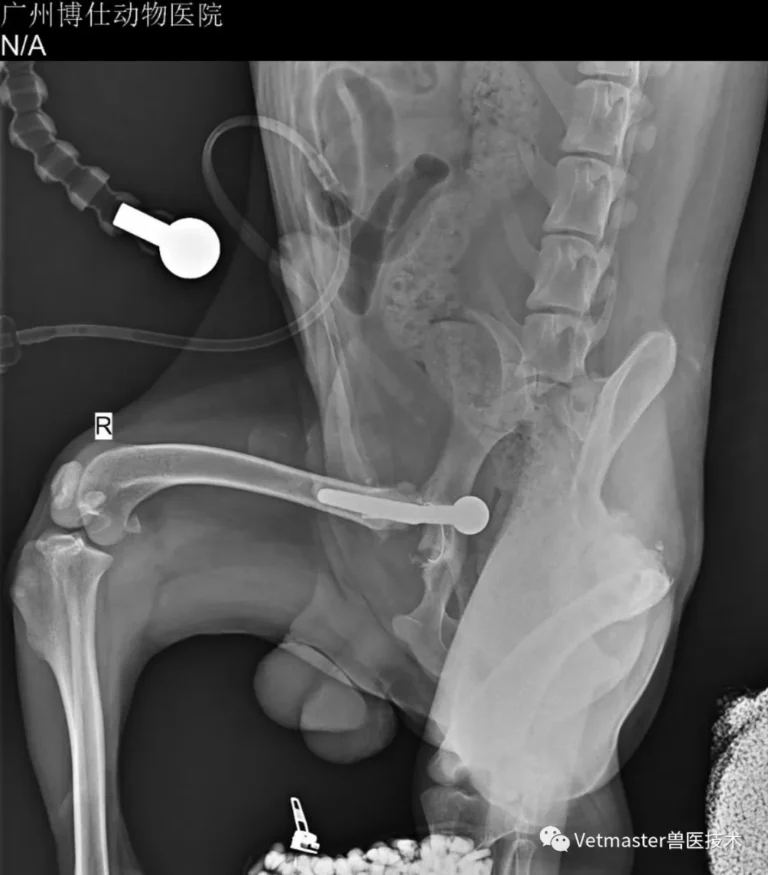
1. Feline Hip Dysplasia: The Hidden Disease
Cats are extraordinary masters at hiding pain. Unlike dogs, who will readily limp or vocalize when a joint hurts, a cat suffering from bilateral hip dysplasia often just quietly withdraws. They stop jumping onto the refrigerator, spend more time sleeping, or adopt a slightly guarded, stiff-legged walk that owners easily misinterpret as laziness or aging.
Consequently, by the time an owner brings a cat to Guangzhou Boss Animal Hospital with overt, visible lameness, the joint is rarely in the early stages. The acetabular cartilage has usually been ground down to eburnated bone, accompanied by chronic periarticular osteophytes.
That was precisely the situation with this patient: a 2-year-old intact male British Shorthair weighing 5.8 kg. His owner noticed a progressive reluctance to move and a visibly altered gait. Physical examination under sedation revealed marked bilateral hip instability, severe discomfort during hip extension, and audible crepitation.
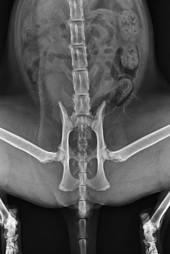
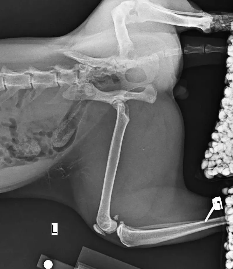
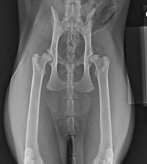
Orthogonal radiographs confirmed symmetrical, advanced bilateral hip dysplasia. The acetabular coverage was severely deficient, the joint margins were ragged with osteophytic rim remodeling, and chronic subluxation had chewed away the articular surfaces. Conservative management had failed to provide lasting relief. For a young, heavy cat with fifteen or more years ahead of him, femoral head and neck ostectomy (FHO) would have left permanent biomechanical deficits and persistent muscle atrophy. The only definitive solution to restore true, pain-free athletic function was total hip replacement (THR).
2. Why Feline THR Demands a Dual-Mobility Biological System
A cat is not just a miniature dog. Feline hip replacements present biological and mechanical demands that rule out standard canine protocols:
- Unique Bone Morphology:
The feline acetabulum is tiny, shallow, and has very little bone stock. Conversely, the feline proximal femur lacks the tapering metaphyseal flare seen in dogs; it is essentially a cylindrical, "stovepipe" canal. - Explosive, "Yoga-Like" Range of Motion:
Cats can jump eight to nine times their body height and routinely twist their hindquarters into extreme physiological angles. A traditional fixed-bearing, single-mobility cup in such a small size has a minimal jump distance (the lateral excursion required before the femoral head pops out of the cup). In an active cat, a standard cup carries an uncomfortably high risk of postoperative luxation. - Longevity & Cementless Fixation:
Because cats frequently live into their late teens or twenties, relying on a thin, 0.5–2 mm mantle of bone cement in a young, active animal is a gamble on aseptic loosening over time. A biological, 3D-printed porous titanium cup and stem that achieve genuine osseointegration are essential for lifelong stability.
For this cat, I selected the Vetmaster cementless dual-mobility hip system: a 12 mm biological press-fit acetabular cup, a #3 straight femoral stem, and a ceramic dual-mobility articulation with a Vitamin E-stabilized highly cross-linked polyethylene liner. The dual-mobility mechanism provides a dramatic increase in effective head diameter during extreme motion, providing the jump distance necessary to absorb feline athletic impacts without popping out.
3. Stage One: Left Total Hip Replacement
I addressed the left hip first.
The primary intraoperative challenge in cats is the vanishingly low margin for error. The feline acetabulum is so small that a deviation of just a few degrees or a millimeter of eccentric reaming will compromise your dorsal rim coverage or risk wall blowout. Furthermore, when applying surgical retractors around such a compact pelvis, any excessive soft-tissue traction easily tilts the entire pelvis on the table, creating a false visual axis for the surgeon.
To counter this, I utilized intraoperative C-arm fluoroscopy to continuously verify the cat's true orthogonal pelvic alignment before and during cup impaction. The 12 mm cup was reamed manually down to bleeding subchondral bone with meticulous care given to preserving the delicate dorsal acetabular rim. The cup was seated at a 45-degree lateral opening angle with physiologic retroversion, achieving an immediate, rock-solid press-fit.
Preparing the stovepipe femur required steady canal broaching to ensure the straight #3 stem achieved uniform axial stability without cortical fracture, ultimately filling over 85% of the proximal medullary canal. After placing a trial reduction, the hip demonstrated full, fluid range of motion without impingement.
The cat recovered smoothly and was bearing comfortable weight on the operated leg within days.
4. Stage Two: Right THR and the Challenge of Contralateral Metal
Five months after the first procedure, the owner brought the cat back. The left hip was performing brilliantly—the limb had regained robust muscle mass, and the cat was moving comfortably. However, with the left leg functioning normally, the chronic pain in the unoperated right hip had become the clear limiting factor in his daily activity. The owner requested that we proceed with the right side.
Replacing the second side, however, introduces a distinct technical hurdle: fluoroscopic artifact and superimposition.
During the first side's surgery, you have an unobstructed lateral fluoroscopic view of the pelvis to confirm the acetabular cup's anteversion and inclination angles. But during the second side, the dense metallic implants in the contralateral left hip project directly across the lateral C-arm field, creating severe visual blockage. You cannot rely on lateral X-rays to directly read the new cup’s orientation during impaction.
Here is how I manage that challenge:
- Pelvic Leveling via C-Arm: Instead of attempting to visualize the new cup through the existing metal, I use the C-arm strictly to verify that the pelvis itself is positioned absolutely flat and neutral relative to the table.
- Mechanical Alignment Rod & Tactile Landmarks: Once the animal's pelvic plane is locked and verified, I orient the new acetabular cup using the mechanical alignment guide rod on the impactor handle, cross-referenced against the anatomical landmarks of the dorsal acetabular rim and ischial spine.
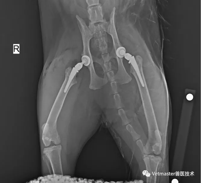
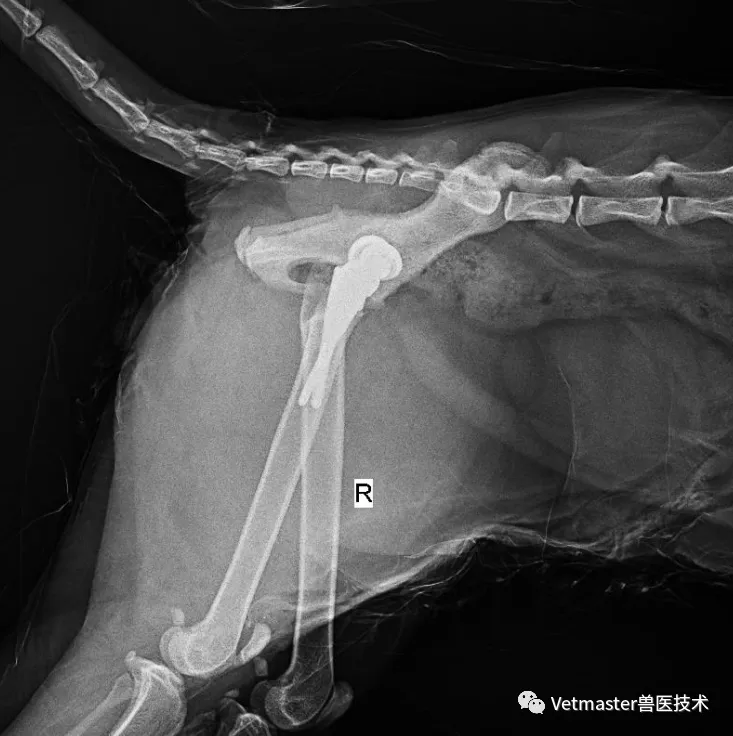
We broached the canal, seated a second #3 stem, and placed a matching 12 mm cup. Postoperative orthogonal radiographs confirmed near-perfect bilateral symmetry: both cups seated securely at approximately 45 degrees of abduction with physiological retroversion, and the femoral stems centered cleanly within their respective canals.
5. Surgical Summary & Key Takeaways
Bilateral hip replacement in a small feline patient is technically demanding, but it is one of the most rewarding procedures in veterinary orthopedics.
Looking back at this case:
- Never Underestimate Feline Joint Pathology: Just because a cat is not crying out doesn't mean his hips aren't ruined. If clinical signs and radiographs show end-stage dysplasia, biological THR provides the same transformative relief in felines as it does in large dogs.
- Dual-Mobility Is a Game Changer for Cats: The combination of an ultra-smooth ceramic head moving inside a mobile UHMWPE bearing gives cats the wide arc of motion their bodies naturally demand, effectively neutralizing the luxation risks that plagued early feline hip surgeries.
- Mastering the Second Side: When performing sequential bilateral replacements, be prepared for the contralateral implant to obscure your lateral fluoroscopy. Train your hands to trust anatomic landmarks and mechanical guide rods once the patient's pelvis has been leveled.
Today, this British Shorthair has two stable, pain-free hips that will serve him for the rest of his life.