1. The French Bulldog Paradox: The Cost of "Cute"
French Bulldogs are among the most anatomically compromised patients to walk through the doors of Guangzhou Boss Animal Hospital. In their pursuit of heavy wrinkles, flat faces, and compact, screw-tailed bodies, human selective breeding has engineered a living catalogue of orthopedic and neurological vulnerabilities:
- The Respiratory Burden: Brachycephalic Airway Obstructive Syndrome (BAOS) leaves them perpetually oxygen-deprived, making every sedation and recovery an exercise in strict airway vigilance.
- The Deformed Vertebral Column: Almost every Frenchie possesses congenital spinal anomalies—butterfly vertebrae, transitional segments, abnormal kyphosis, and hemivertebrae.
- The Dysplastic Pelvis: Their hips are notoriously shallow, with high rates of congenital dysplasia and early degenerative remodeling.
When a breed carries this many overlapping structural flaws, pinpointing why a dog has suddenly stopped walking requires a surgeon to look past superficial radiographic signs and cut straight to the physiological root cause.
2. Case Presentation: Blaming the Hips for a Paralyzed Spine
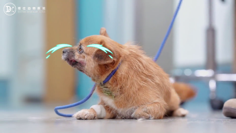
The patient was an adult French Bulldog brought in for progressive "hindlimb weakness."
His owners had taken him to an outside practice where radiographs showed shallow acetabula, leading the attending vet to diagnose hip dysplasia. They tried conservative rest, but the episodes grew increasingly frequent and severe until the dog was dragging his hindquarters.
The moment he was placed on my exam table, the true diagnosis was obvious:
- Neurological Deficits: The dog had significant conscious proprioceptive deficits, primarily in the left hindlimb, delayed knuckling correction, and upper motor neuron paraparesis.
- The Clinical Rule: As I have stated countless times in my clinical writing: canine hip dysplasia does not cause paralysis. A dysplastic hip hurts, causes a bunny-hopping gait, and produces disuse atrophy; it does not abolish spinal reflexes or erase proprioception. If a dog is dragging its paws and stumbling neurological steps, the emergency is in the spine, not the pelvis.
Conservative therapy had reached its limits. We moved the dog immediately into our imaging suite for high-resolution computed tomography (CT).
3. CT Analysis: Peeling Back the Anatomical Layers
CT scanning in a French Bulldog is an exercise in sorting incidental anomalies from surgical emergencies:
- The Surgical Culprit (T13-L1):
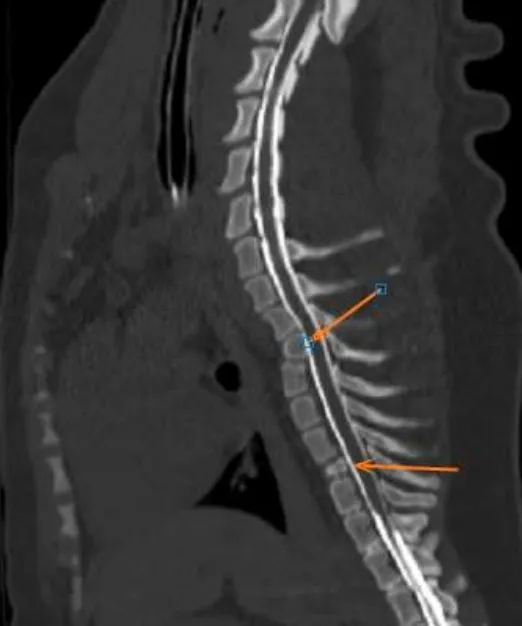
Between the thirteenth thoracic and first lumbar vertebrae, an acute extruded intervertebral disc herniation occupied the ventral and ventrolateral spinal canal. The mineralized disc material was impinging on the thecal sac, resulting in 30% to 40% spinal cord compression. This was the single, direct cause of his paraparesis. - Incidental Congenital Deformities (T4 & T8):
Further up the thoracic column, our 3D reconstructions revealed classic wedge-shaped hemivertebrae at T4 and T8, creating an irregular regional kyphoscoliosis. However, there was no active canal stenosis or disc extrusion at these sites. Operating on these incidental birth defects would have been entirely inappropriate.
- The Incidental Hips:
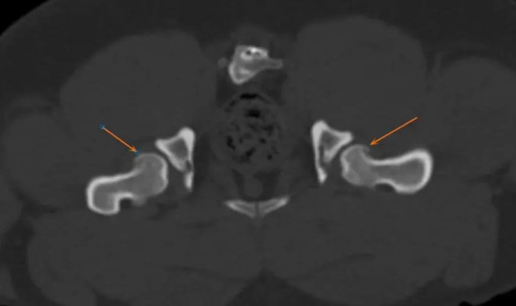
Examining his pelvic scans confirmed what the outside clinic had seen: bilaterally shallow acetabula with subluxated femoral heads and mild periarticular osteophytes. But again: this chronic hip dysplasia was entirely incidental to his acute paralysis.
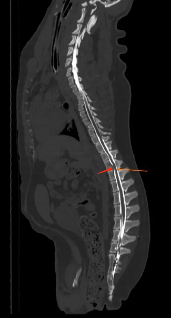
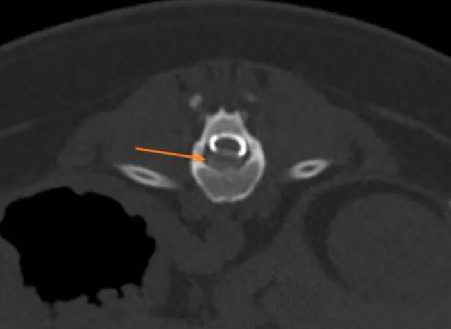
My objective was clear: perform an emergency spinal decompression at T13-L1 and leave the rest of his anatomy alone.
4. Surgical Decompression: T13-L1 Hemilaminectomy
Under continuous multimodal anesthesia and rigid respiratory monitoring, I took the dog to our clean-room surgical suite.
- Exposure:
Approaching from the left dorsolateral aspect of T13-L1, I carefully elevated the epaxial musculature, identifying the precise neurovascular and bony landmarks of the articular facets and pedicles. - Window Creation:
Using a high-speed surgical pneumatic drill with continuous chilled irrigation, I burred away the articular facets and thinned the pedicles of T13 and L1. - Decompression:
Once the inner cortical layer was paper-thin, it was elevated to expose the compressed thecal sac. The extruded, chalky disc material lay wedged beneath the cord. Working under microsurgical magnification with micro-curettes, I carefully teased out the herniated fragments until the dura relaxed, epidural venous pulsations returned, and the entire ventrolateral canal was unobstructed. - Closure:
A protective autologous fat graft was placed over the laminectomy defect, followed by anatomical layer-by-layer closure of the muscular fascia and skin.
5. Recovery: "Never Forgetting the Food Bowl"
If there is one thing I deeply admire about French Bulldogs, it is their indomitable spirit.
Spinal surgery is an intensive procedure, but the moment this little guy cleared anesthesia and his extubation was safely confirmed, his lethargy vanished. As I captured in my clinical notes that evening: he woke up from spinal surgery and immediately dove headfirst into his food bowl without missing a beat.
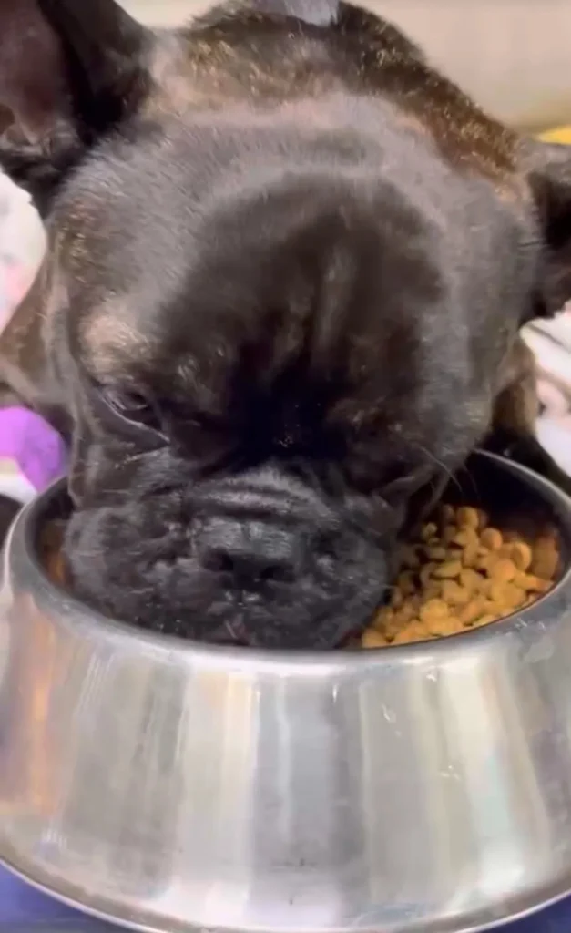
His recovery followed a steady trajectory:
- Left hindlimb proprioception began recovering within 72 hours.
- Supported by strict crate rest for the first four weeks, followed by controlled physical therapy, his motor strength returned rapidly.
- Within a few weeks, he had completely regained unassisted, coordinated four-legged ambulation. The dragging was eliminated, spinal pain had vanished, and his overall quality of life was fully restored.
6. Clinical Reflections from the Operating Theater
This case encapsulates two core truths that I drill into interns and visiting veterinarians at our hospital:
- "See Through the Surface to Find the Truth":
Never diagnose based on a single abnormal radiograph. In brachycephalic and chondrodystrophic breeds, abnormal X-rays are the baseline. If you operate on every shallow hip or wedge vertebra you see, you will subject animals to pointless surgeries while their actual life-threatening disc herniations continue to strangle their spinal cords. Always let the neurological examination guide your knife. - Welfare Beyond the Clinic:
As surgeons, we can drill windows into spines and stabilize wrecked joints, but we must never normalize these deformities. Hemivertebrae, collapsed nostrils, elongated soft palates, and deformed hips are not "cute breed quirks"—they are physical handicaps created by human aesthetics. While we dedicate our careers to fixing them on the operating table, the veterinary profession must continue to demand responsible breeding that puts anatomical health above cosmetic exaggeration.
For this Frenchie, an accurate diagnosis saved him from unnecessary hip interventions and gave him back his legs—and true to form, he celebrated every single day of it at the dinner bowl.