
1. Introduction
Total knee replacement in small animals is relatively rare, primarily due to the stringent requirements for prosthetic precision and the lack of specialized surgical instrumentation. The integration of 3D printing technology has enabled the creation of custom, high-precision implants. This case demonstrates the clinical efficacy of custom 3D-printed implants developed by Dr. Wu Zhongheng’s team in collaboration with materials science experts to address complex joint pathology.
2. Case Presentation
2.1 Patient Profile and Clinical Signs
The patient was a 25kg Husky presenting with significant lameness in the left hind limb. Physical examination revealed crepitus during knee flexion and extension, a positive tibial compression test, and ambiguous results from the drawer test. These clinical signs indicated meniscal injury secondary to a chronic cranial cruciate ligament tear, which had progressed into severe osteoarthritis.
2.2 Diagnostic Imaging
Radiographic and CT evaluations confirmed the physical exam findings, showing irregular joint margins and severe degenerative changes. Concurrent examination of the hip joints showed no abnormalities, isolating the pathology to the knee joint.
3. Preoperative Planning and Implant Technology
3.1 Development of 3D-Printed Implants
The surgical team established a strategic partnership with the Institute of New Materials at the Guangdong Academy of Sciences to form a specialized medical-engineering group.

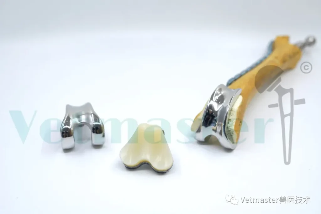
The implants were produced via 3D metal printing and consisted of a femoral condyle prosthesis, a tibial plateau prosthesis, and a meniscal insert. The system utilized a composite structure of CoCrMo alloy, porous titanium alloy, and Vitamin E cross-linked polyethylene; a technical search by the Ministry of Science and Technology confirmed this as the world’s first report of a canine TKR utilizing this specific material combination. All materials underwent toxicological and mechanical testing to ensure they met implant-grade safety standards.

3.2 Surgical Simulation and Custom Tooling
Prior to the actual surgery, a 1:1 scale plastic model of the patient’s skeleton was created using 3D printing. The surgeons performed multiple step-by-step simulations on this model to confirm the fit between the implants and the bone, and to verify the performance of the custom 3D-printed disposable surgical tools, thereby minimizing unpredictable intraoperative risks.
4. Surgical Results and Follow-up Assessment
4.1 Immediate Postoperative Recovery
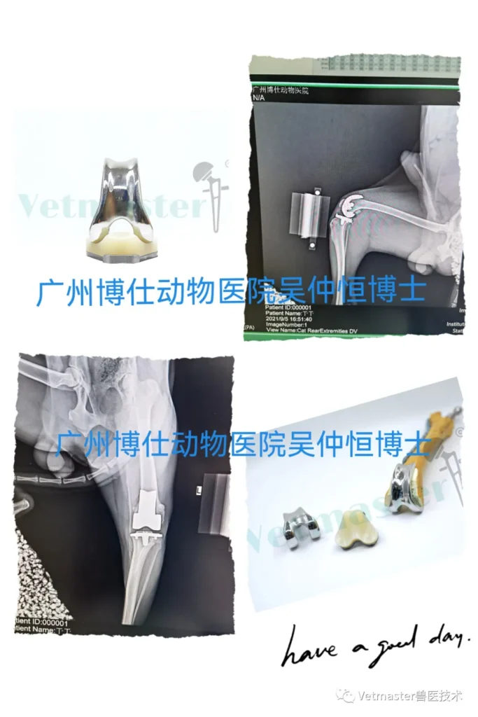
Intraoperatively, the implants were stable and achieved a perfect anatomical fit with the patient’s bone. By the first day following surgery, the dog showed excellent tolerance and was able to engage in pain-free walking.
4.2 Long-term Efficacy Assessment
Follow-up at 2.5 months post-surgery showed outstanding recovery. Video footage recorded the Husky running freely and joyfully on grass, with a gait showing no signs of having undergone major surgery. While the recovery cycle for TKR is longer than that of hip replacement, the complication rate is reported to be lower.
5. Conclusion
This case proves the feasibility of custom 3D printing technology in small animal clinical practice. For knee joints with irreversible damage and severe arthritis, custom total knee replacement serves as an effective ultimate solution for restoring mobility and enhancing quality of life.