
1. Case Background
1.1 Initial Diagnosis and Surgical History
The patient, a Shiba Inu, was diagnosed with hip dysplasia 10 months prior during an initial consultation at Guangzhou Boshi Animal Hospital. Subsequently, the patient underwent a left-side cemented THR at another clinic. However, approximately eight months post-surgery, the dog exhibited recurrent pain, leading to a request for specialized revision.
2. Diagnostic Evaluation
2.1 Radiographic Feature Analysis
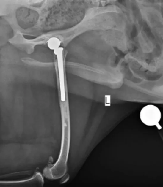
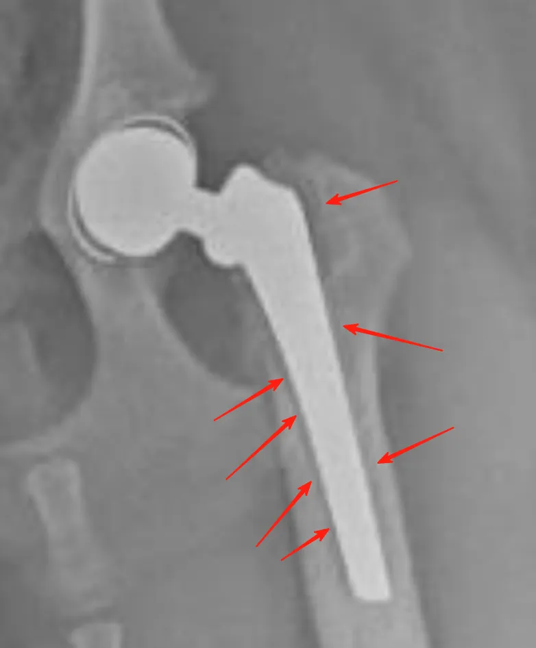
Radiographs revealed a prominent radiolucent zone surrounding the femoral stem, confirming the loosening of the cemented implant. Comparison with earlier images showed a clear posterior rotation and displacement of the stem.
2.2 Secondary Pathological Changes
Further observation identified a periosteal reaction along the medial aspect of the femur, accompanied by resorption of the medial cortical bone. these signs collectively indicated a failure of the cement-bone interface leading to biomechanical instability.
3. Surgical Revision Procedure
3.1 Removal of Failed Implants
The primary surgical objective was the removal of the loosened cemented components. The stem and proximal cement were successfully extracted. Due to the excessive depth of some cement within the medullary canal, a small amount of distal cement was left in situ to avoid the risk of secondary fractures during retrieval.
3.2 Implantation of biological Components
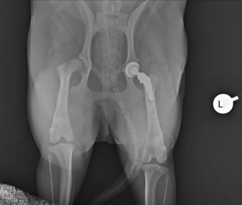
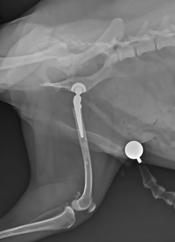
Following debridement, the team utilized a Vetmaster biological dual-mobility hip system for the revision. Despite the compromised osseointegration capacity caused by previous cement exposure, precise surgical maneuvering achieved excellent press-fit stability for the new prosthesis.
4. Postoperative Follow-up and Rehabilitation
4.1 Radiographic Re-evaluation
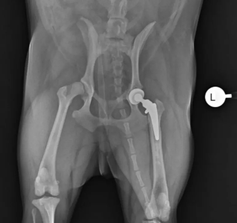
Follow-up at two months post-revision showed that the biological implants remained stable. Radiographic evidence of early bone ingrowth was observed in several areas. The distal residual cement did not interfere with the stability of the new prosthesis.
4.2 Functional Recovery
The patient’s mobility returned to ideal levels, with a complete resolution of pain. Measurements indicated that the muscle diameter of the operated limb had recovered to match, or even slightly exceed, the contralateral limb, signifying full functional reconstruction.
5. Conclusion
Cemented total hip replacements carry a long-term risk of implant loosening in certain cases. For such failures, biological dual-mobility systems provide a robust revision alternative. By maximizing the contact area and utilizing biological fixation, clinicians can effectively resolve pain associated with loosening and restore the patient's quality of life.