1. Case Presentation
The patient was a 2-year-old male British Shorthair cat weighing 5.8 kg. The owner reported a significant decrease in daily activity and an abnormal gait. Physical examination revealed pronounced pain responses in both hip joints and restricted physiological range of motion.
2. Imaging Evaluation
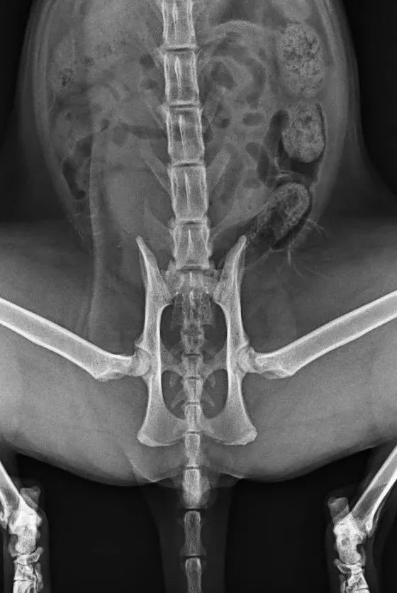
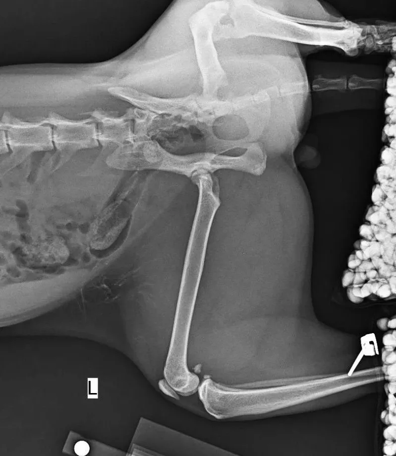
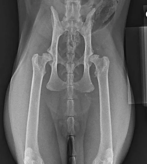
Preoperative radiographs confirmed symmetrical bilateral hip dysplasia. The primary pathological features included a significant reduction in acetabular coverage of the femoral head, accompanied by marginal osteophyte formation and subchondral bone wear. The chronic mechanical instability of the joints had led to the rapid progression of degenerative osteoarthritis.
3. Surgical Planning and Implant Selection
Considering the long life expectancy and the explosive jumping ability of felines, a biological (cementless) dual-mobility total hip system was selected. This system utilizes a microporous surface for bone ingrowth to achieve long-term biological fixation. Specific implant sizes included a 12mm acetabular cup, a #3 femoral stem, and a compatible ceramic dual-mobility head. The dual-mobility design was chosen to provide an increased range of motion and minimize the risk of postoperative luxation.
4. Surgical Procedure and Technical Challenges
4.1 Stage One: Left THR
The medical team first operated on the left limb. Because the feline acetabulum is extremely small, the margin for error is low. Any positional shift caused by instrument tension during reaming could compromise the implantation angle. Intraoperative C-arm fluoroscopy was used to repeatedly verify the animal's positioning, ensuring that the acetabular cup's abduction and anteversion angles aligned with anatomical standards. Given the "stovepipe" (cylindrical) shape of the feline proximal femur, precise centralization of the #3 stem was critical to occupy over 85% of the medullary canal.
4.2 Stage Two: Right THR
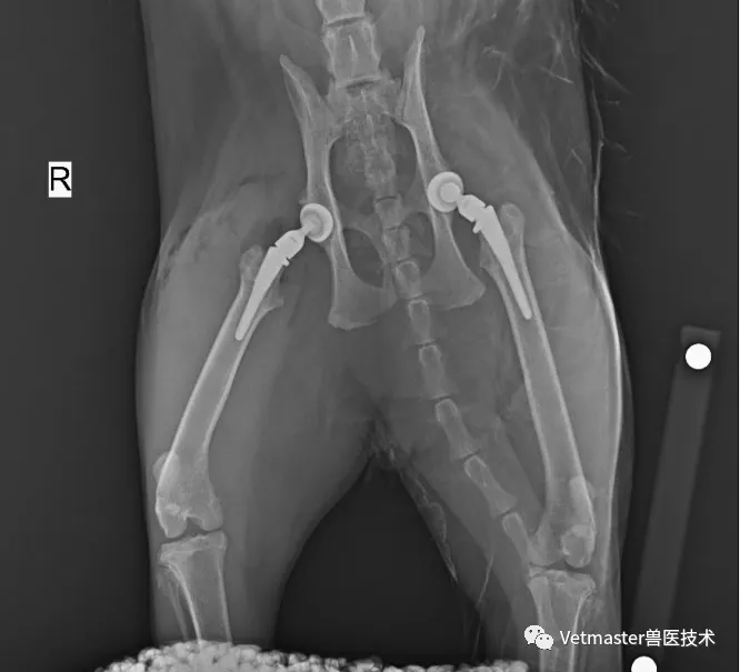
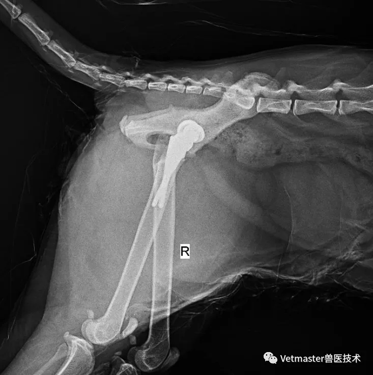
Five months after the initial surgery, following excellent weight-bearing and mobility recovery of the left limb, the right hip was replaced. The presence of the metal implant on the contralateral side caused some interference during imaging. The surgeon utilized a combination of C-arm monitoring and reamer alignment guides to achieve precise prosthesis positioning once again.
5. Discussion and Conclusion
Feline THR is anatomically unique; the acetabular-to-femoral ratio and the medullary canal morphology differ significantly from those in canines. Technical challenges include high-precision maneuvers in small joints and maintaining intraoperative positioning. This case successfully resolved chronic pain through staged bilateral replacement. Clinical outcomes suggest that for highly active felines, utilizing a dual-mobility system with superior anti-dislocation properties combined with precise image guidance is an effective ultimate solution for severe joint pathology.